Manganese »
PDB 2who-2ypp »
2ygk »
Manganese in PDB 2ygk: Crystal Structure of the Nura Nuclease From Sulfolobus Solfataricus
Protein crystallography data
The structure of Crystal Structure of the Nura Nuclease From Sulfolobus Solfataricus, PDB code: 2ygk
was solved by
N.J.Rzechorzek,
J.K.Blackwood,
A.S.Abrams,
J.D.Maman,
N.P.Robinson,
L.Pellegrini,
with X-Ray Crystallography technique. A brief refinement statistics is given in the table below:
| Resolution Low / High (Å) | 35.94 / 2.50 |
| Space group | P 1 21 1 |
| Cell size a, b, c (Å), α, β, γ (°) | 67.740, 80.280, 67.670, 90.00, 107.17, 90.00 |
| R / Rfree (%) | 23.176 / 27.089 |
Manganese Binding Sites:
The binding sites of Manganese atom in the Crystal Structure of the Nura Nuclease From Sulfolobus Solfataricus
(pdb code 2ygk). This binding sites where shown within
5.0 Angstroms radius around Manganese atom.
In total 2 binding sites of Manganese where determined in the Crystal Structure of the Nura Nuclease From Sulfolobus Solfataricus, PDB code: 2ygk:
Jump to Manganese binding site number: 1; 2;
In total 2 binding sites of Manganese where determined in the Crystal Structure of the Nura Nuclease From Sulfolobus Solfataricus, PDB code: 2ygk:
Jump to Manganese binding site number: 1; 2;
Manganese binding site 1 out of 2 in 2ygk
Go back to
Manganese binding site 1 out
of 2 in the Crystal Structure of the Nura Nuclease From Sulfolobus Solfataricus
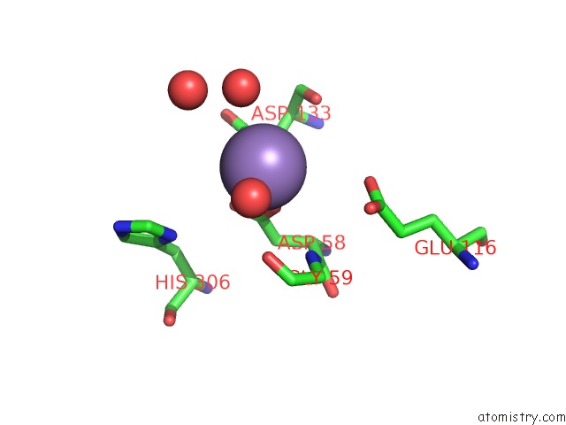
Mono view
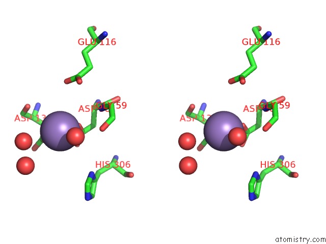
Stereo pair view
Mono view
Stereo pair view
A full contact list of Manganese with other atoms in the Mn binding
site number 1 of Crystal Structure of the Nura Nuclease From Sulfolobus Solfataricus within 5.0Å range:
|
Manganese binding site 2 out of 2 in 2ygk
Go back to
Manganese binding site 2 out
of 2 in the Crystal Structure of the Nura Nuclease From Sulfolobus Solfataricus
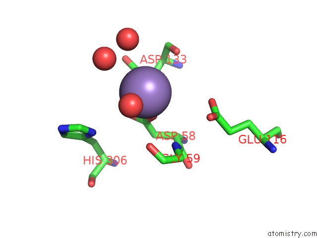
Mono view
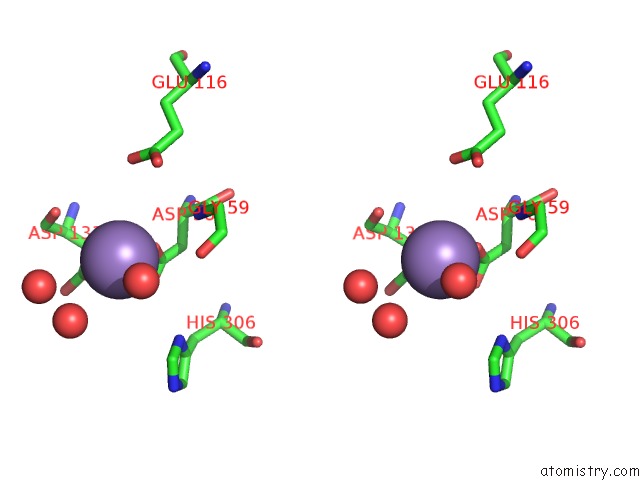
Stereo pair view
Mono view
Stereo pair view
A full contact list of Manganese with other atoms in the Mn binding
site number 2 of Crystal Structure of the Nura Nuclease From Sulfolobus Solfataricus within 5.0Å range:
|
Reference:
J.K.Blackwood,
N.J.Rzechorzek,
A.S.Abrams,
J.D.Maman,
L.Pellegrini,
N.P.Robinson.
Structural and Functional Insights Into Dna-End Processing By the Archaeal Hera Helicase-Nura Nuclease Complex. Nucleic Acids Res. V. 40 3183 2012.
ISSN: ISSN 0305-1048
PubMed: 22135300
DOI: 10.1093/NAR/GKR1157
Page generated: Sat Oct 5 15:31:52 2024
ISSN: ISSN 0305-1048
PubMed: 22135300
DOI: 10.1093/NAR/GKR1157
Last articles
Zn in 9J0NZn in 9J0O
Zn in 9J0P
Zn in 9FJX
Zn in 9EKB
Zn in 9C0F
Zn in 9CAH
Zn in 9CH0
Zn in 9CH3
Zn in 9CH1