Manganese »
PDB 2who-2ypp »
2wjd »
Manganese in PDB 2wjd: Crystal Structure of the Tyrosine Phosphatase CPS4B From Steptococcus Pneumoniae TIGR4.
Enzymatic activity of Crystal Structure of the Tyrosine Phosphatase CPS4B From Steptococcus Pneumoniae TIGR4.
All present enzymatic activity of Crystal Structure of the Tyrosine Phosphatase CPS4B From Steptococcus Pneumoniae TIGR4.:
3.1.3.48;
3.1.3.48;
Protein crystallography data
The structure of Crystal Structure of the Tyrosine Phosphatase CPS4B From Steptococcus Pneumoniae TIGR4., PDB code: 2wjd
was solved by
G.Hagelueken,
H.Huang,
J.H.Naismith,
with X-Ray Crystallography technique. A brief refinement statistics is given in the table below:
| Resolution Low / High (Å) | 29.08 / 2.80 |
| Space group | P 43 21 2 |
| Cell size a, b, c (Å), α, β, γ (°) | 88.850, 88.850, 92.310, 90.00, 90.00, 90.00 |
| R / Rfree (%) | 17.3 / 23.2 |
Other elements in 2wjd:
The structure of Crystal Structure of the Tyrosine Phosphatase CPS4B From Steptococcus Pneumoniae TIGR4. also contains other interesting chemical elements:
| Samarium | (Sm) | 2 atoms |
Manganese Binding Sites:
The binding sites of Manganese atom in the Crystal Structure of the Tyrosine Phosphatase CPS4B From Steptococcus Pneumoniae TIGR4.
(pdb code 2wjd). This binding sites where shown within
5.0 Angstroms radius around Manganese atom.
In total 3 binding sites of Manganese where determined in the Crystal Structure of the Tyrosine Phosphatase CPS4B From Steptococcus Pneumoniae TIGR4., PDB code: 2wjd:
Jump to Manganese binding site number: 1; 2; 3;
In total 3 binding sites of Manganese where determined in the Crystal Structure of the Tyrosine Phosphatase CPS4B From Steptococcus Pneumoniae TIGR4., PDB code: 2wjd:
Jump to Manganese binding site number: 1; 2; 3;
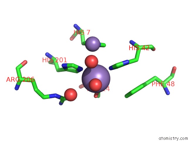
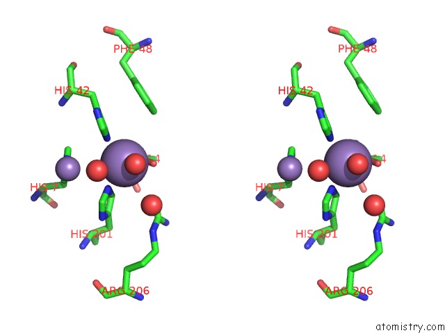
Manganese binding site 1 out of 3 in 2wjd
Go back to
Manganese binding site 1 out
of 3 in the Crystal Structure of the Tyrosine Phosphatase CPS4B From Steptococcus Pneumoniae TIGR4.
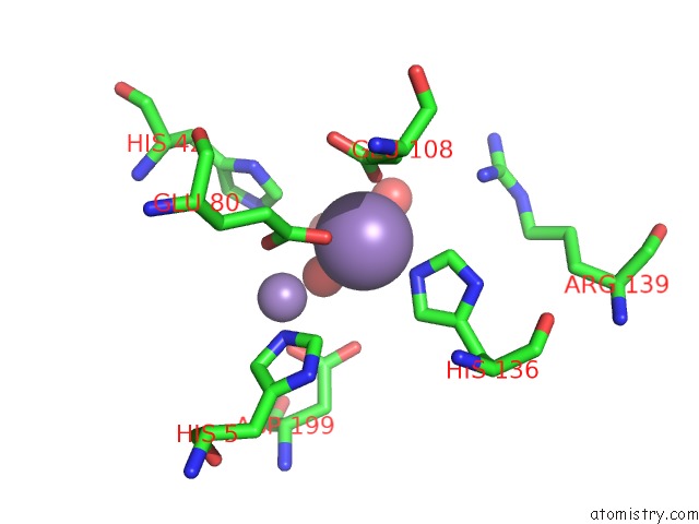
Mono view
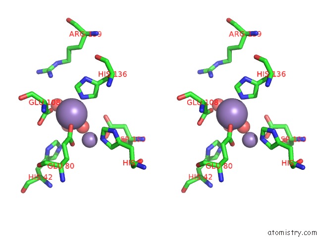
Stereo pair view
Mono view
Stereo pair view
A full contact list of Manganese with other atoms in the Mn binding
site number 1 of Crystal Structure of the Tyrosine Phosphatase CPS4B From Steptococcus Pneumoniae TIGR4. within 5.0Å range:
|
Manganese binding site 2 out of 3 in 2wjd
Go back to
Manganese binding site 2 out
of 3 in the Crystal Structure of the Tyrosine Phosphatase CPS4B From Steptococcus Pneumoniae TIGR4.
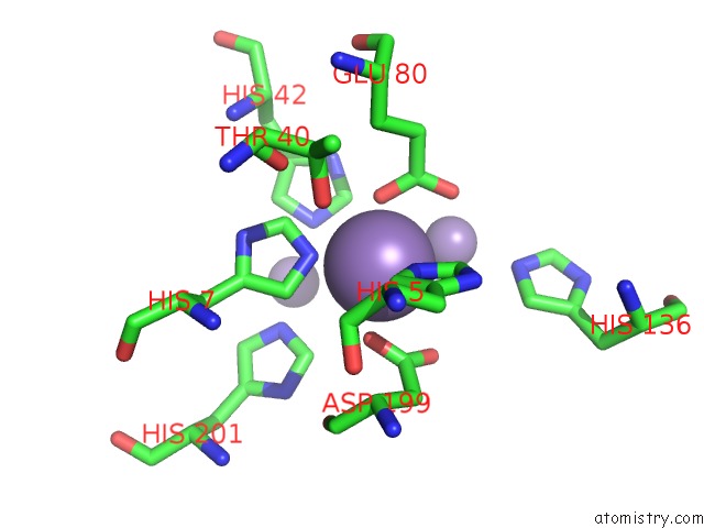
Mono view
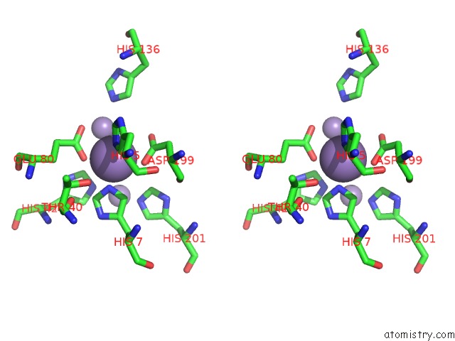
Stereo pair view
Mono view
Stereo pair view
A full contact list of Manganese with other atoms in the Mn binding
site number 2 of Crystal Structure of the Tyrosine Phosphatase CPS4B From Steptococcus Pneumoniae TIGR4. within 5.0Å range:
|
Manganese binding site 3 out of 3 in 2wjd
Go back to
Manganese binding site 3 out
of 3 in the Crystal Structure of the Tyrosine Phosphatase CPS4B From Steptococcus Pneumoniae TIGR4.
Mono view
Stereo pair view
Mono view
Stereo pair view
A full contact list of Manganese with other atoms in the Mn binding
site number 3 of Crystal Structure of the Tyrosine Phosphatase CPS4B From Steptococcus Pneumoniae TIGR4. within 5.0Å range:
|
Reference:
G.Hagelueken,
H.Huang,
I.L.Mainprize,
C.Whitfield,
J.H.Naismith.
Crystal Structures of Wzb of Escherichia Coli and Cpsb of Streptococcus Pneumoniae, Representatives of Two Families of Tyrosine Phosphatases That Regulate Capsule Assembly. J.Mol.Biol. V. 392 678 2009.
ISSN: ISSN 0022-2836
PubMed: 19616007
DOI: 10.1016/J.JMB.2009.07.026
Page generated: Sat Oct 5 15:23:52 2024
ISSN: ISSN 0022-2836
PubMed: 19616007
DOI: 10.1016/J.JMB.2009.07.026
Last articles
Fe in 2YXOFe in 2YRS
Fe in 2YXC
Fe in 2YNM
Fe in 2YVJ
Fe in 2YP1
Fe in 2YU2
Fe in 2YU1
Fe in 2YQB
Fe in 2YOO