Manganese »
PDB 2v3z-2wgz »
2w42 »
Manganese in PDB 2w42: The Structure of A Piwi Protein From Archaeoglobus Fulgidus Complexed with A 16NT Dna Duplex.
Protein crystallography data
The structure of The Structure of A Piwi Protein From Archaeoglobus Fulgidus Complexed with A 16NT Dna Duplex., PDB code: 2w42
was solved by
J.S.Parker,
S.M.Roe,
D.Barford,
with X-Ray Crystallography technique. A brief refinement statistics is given in the table below:
| Resolution Low / High (Å) | 98.06 / 1.90 |
| Space group | P 1 |
| Cell size a, b, c (Å), α, β, γ (°) | 51.838, 61.384, 103.540, 75.80, 75.86, 79.47 |
| R / Rfree (%) | 20.5 / 24.3 |
Manganese Binding Sites:
The binding sites of Manganese atom in the The Structure of A Piwi Protein From Archaeoglobus Fulgidus Complexed with A 16NT Dna Duplex.
(pdb code 2w42). This binding sites where shown within
5.0 Angstroms radius around Manganese atom.
In total 2 binding sites of Manganese where determined in the The Structure of A Piwi Protein From Archaeoglobus Fulgidus Complexed with A 16NT Dna Duplex., PDB code: 2w42:
Jump to Manganese binding site number: 1; 2;
In total 2 binding sites of Manganese where determined in the The Structure of A Piwi Protein From Archaeoglobus Fulgidus Complexed with A 16NT Dna Duplex., PDB code: 2w42:
Jump to Manganese binding site number: 1; 2;
Manganese binding site 1 out of 2 in 2w42
Go back to
Manganese binding site 1 out
of 2 in the The Structure of A Piwi Protein From Archaeoglobus Fulgidus Complexed with A 16NT Dna Duplex.
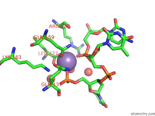
Mono view
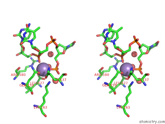
Stereo pair view
Mono view
Stereo pair view
A full contact list of Manganese with other atoms in the Mn binding
site number 1 of The Structure of A Piwi Protein From Archaeoglobus Fulgidus Complexed with A 16NT Dna Duplex. within 5.0Å range:
|
Manganese binding site 2 out of 2 in 2w42
Go back to
Manganese binding site 2 out
of 2 in the The Structure of A Piwi Protein From Archaeoglobus Fulgidus Complexed with A 16NT Dna Duplex.
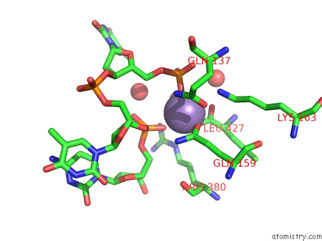
Mono view
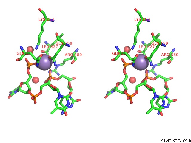
Stereo pair view
Mono view
Stereo pair view
A full contact list of Manganese with other atoms in the Mn binding
site number 2 of The Structure of A Piwi Protein From Archaeoglobus Fulgidus Complexed with A 16NT Dna Duplex. within 5.0Å range:
|
Reference:
J.S.Parker,
E.A.Parizotto,
M.Wang,
S.M.Roe,
D.Barford.
Enhancement of the Seed-Target Recognition Step in Rna Silencing By A Piwi-Mid Domain Protein Mol.Cell V. 33 204 2009.
ISSN: ISSN 1097-2765
PubMed: 19187762
DOI: 10.1016/J.MOLCEL.2008.12.012
Page generated: Sat Oct 5 15:20:41 2024
ISSN: ISSN 1097-2765
PubMed: 19187762
DOI: 10.1016/J.MOLCEL.2008.12.012
Last articles
Cl in 5VQTCl in 5VPF
Cl in 5VPD
Cl in 5VPE
Cl in 5VPC
Cl in 5VOO
Cl in 5VP5
Cl in 5VOH
Cl in 5VPA
Cl in 5VOF