Manganese »
PDB 2v3z-2wgz »
2vqa »
Manganese in PDB 2vqa: Protein-Folding Location Can Regulate Mn Versus Cu- or Zn- Binding. Crystal Structure of Mnca.
Protein crystallography data
The structure of Protein-Folding Location Can Regulate Mn Versus Cu- or Zn- Binding. Crystal Structure of Mnca., PDB code: 2vqa
was solved by
S.Tottey,
K.J.Waldron,
S.J.Firbank,
B.Reale,
C.Bessant,
K.Sato,
J.Gray,
M.J.Banfield,
C.Dennison,
N.J.Robinson,
with X-Ray Crystallography technique. A brief refinement statistics is given in the table below:
| Resolution Low / High (Å) | 58.32 / 2.95 |
| Space group | P 65 2 2 |
| Cell size a, b, c (Å), α, β, γ (°) | 236.190, 236.190, 134.041, 90.00, 90.00, 120.00 |
| R / Rfree (%) | 19.1 / 23.3 |
Manganese Binding Sites:
The binding sites of Manganese atom in the Protein-Folding Location Can Regulate Mn Versus Cu- or Zn- Binding. Crystal Structure of Mnca.
(pdb code 2vqa). This binding sites where shown within
5.0 Angstroms radius around Manganese atom.
In total 6 binding sites of Manganese where determined in the Protein-Folding Location Can Regulate Mn Versus Cu- or Zn- Binding. Crystal Structure of Mnca., PDB code: 2vqa:
Jump to Manganese binding site number: 1; 2; 3; 4; 5; 6;
In total 6 binding sites of Manganese where determined in the Protein-Folding Location Can Regulate Mn Versus Cu- or Zn- Binding. Crystal Structure of Mnca., PDB code: 2vqa:
Jump to Manganese binding site number: 1; 2; 3; 4; 5; 6;
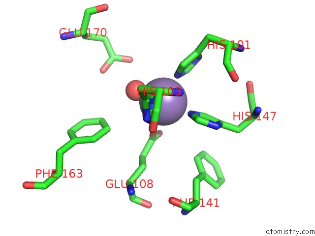
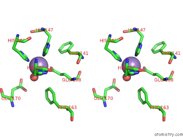
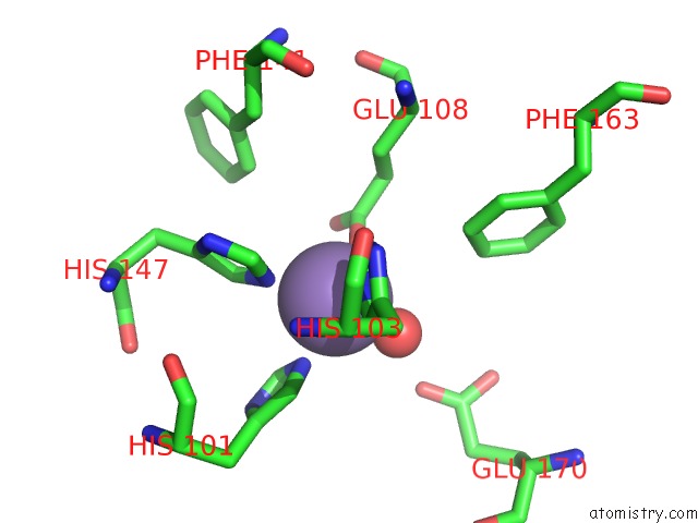
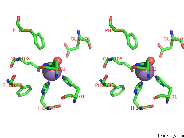
Manganese binding site 1 out of 6 in 2vqa
Go back to
Manganese binding site 1 out
of 6 in the Protein-Folding Location Can Regulate Mn Versus Cu- or Zn- Binding. Crystal Structure of Mnca.
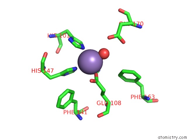
Mono view
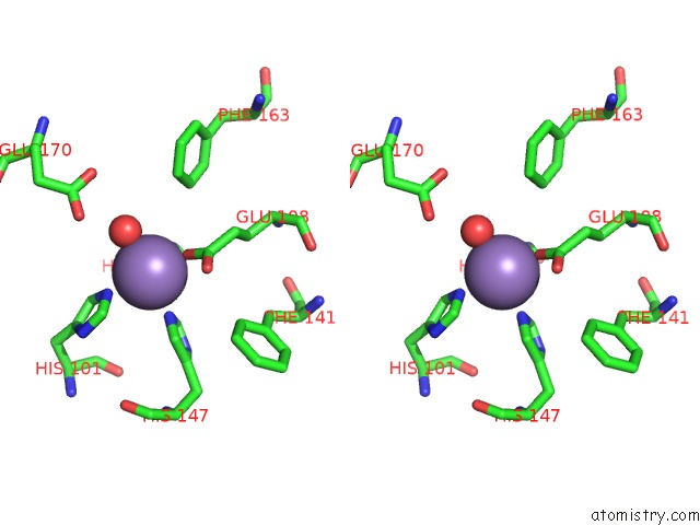
Stereo pair view
Mono view
Stereo pair view
A full contact list of Manganese with other atoms in the Mn binding
site number 1 of Protein-Folding Location Can Regulate Mn Versus Cu- or Zn- Binding. Crystal Structure of Mnca. within 5.0Å range:
|
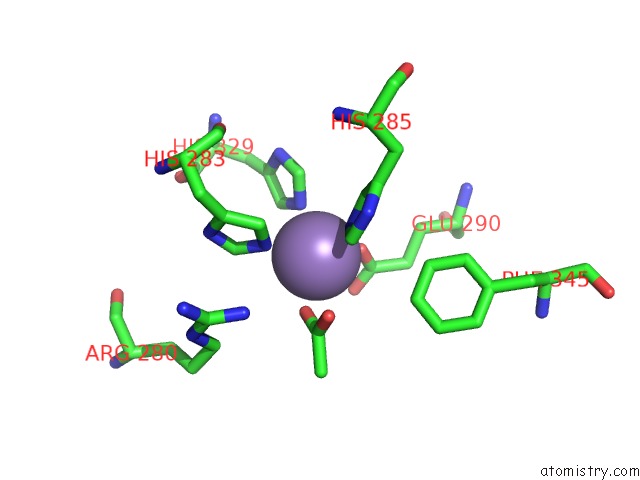
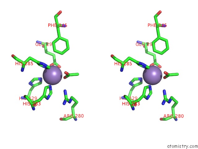
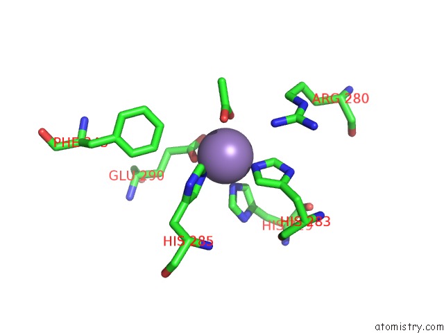
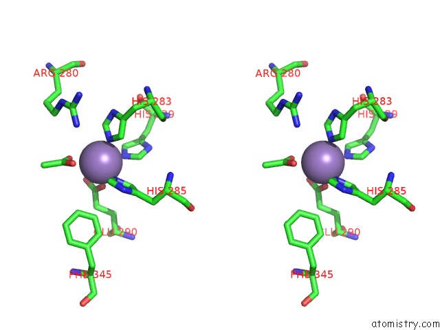
Manganese binding site 2 out of 6 in 2vqa
Go back to
Manganese binding site 2 out
of 6 in the Protein-Folding Location Can Regulate Mn Versus Cu- or Zn- Binding. Crystal Structure of Mnca.
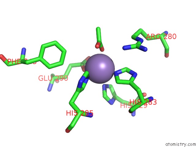
Mono view
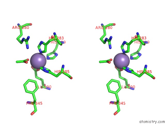
Stereo pair view
Mono view
Stereo pair view
A full contact list of Manganese with other atoms in the Mn binding
site number 2 of Protein-Folding Location Can Regulate Mn Versus Cu- or Zn- Binding. Crystal Structure of Mnca. within 5.0Å range:
|
Manganese binding site 3 out of 6 in 2vqa
Go back to
Manganese binding site 3 out
of 6 in the Protein-Folding Location Can Regulate Mn Versus Cu- or Zn- Binding. Crystal Structure of Mnca.
Mono view
Stereo pair view
Mono view
Stereo pair view
A full contact list of Manganese with other atoms in the Mn binding
site number 3 of Protein-Folding Location Can Regulate Mn Versus Cu- or Zn- Binding. Crystal Structure of Mnca. within 5.0Å range:
|
Manganese binding site 4 out of 6 in 2vqa
Go back to
Manganese binding site 4 out
of 6 in the Protein-Folding Location Can Regulate Mn Versus Cu- or Zn- Binding. Crystal Structure of Mnca.
Mono view
Stereo pair view
Mono view
Stereo pair view
A full contact list of Manganese with other atoms in the Mn binding
site number 4 of Protein-Folding Location Can Regulate Mn Versus Cu- or Zn- Binding. Crystal Structure of Mnca. within 5.0Å range:
|
Manganese binding site 5 out of 6 in 2vqa
Go back to
Manganese binding site 5 out
of 6 in the Protein-Folding Location Can Regulate Mn Versus Cu- or Zn- Binding. Crystal Structure of Mnca.
Mono view
Stereo pair view
Mono view
Stereo pair view
A full contact list of Manganese with other atoms in the Mn binding
site number 5 of Protein-Folding Location Can Regulate Mn Versus Cu- or Zn- Binding. Crystal Structure of Mnca. within 5.0Å range:
|
Manganese binding site 6 out of 6 in 2vqa
Go back to
Manganese binding site 6 out
of 6 in the Protein-Folding Location Can Regulate Mn Versus Cu- or Zn- Binding. Crystal Structure of Mnca.
Mono view
Stereo pair view
Mono view
Stereo pair view
A full contact list of Manganese with other atoms in the Mn binding
site number 6 of Protein-Folding Location Can Regulate Mn Versus Cu- or Zn- Binding. Crystal Structure of Mnca. within 5.0Å range:
|
Reference:
S.Tottey,
K.J.Waldron,
S.J.Firbank,
B.Reale,
C.Bessant,
K.Sato,
T.R.Cheek,
J.Gray,
M.J.Banfield,
C.Dennison,
N.J.Robinson.
Protein-Folding Location Can Regulate Manganese-Binding Versus Copper- or Zinc-Binding. Nature V. 455 1138 2008.
ISSN: ISSN 0028-0836
PubMed: 18948958
DOI: 10.1038/NATURE07340
Page generated: Sat Oct 5 15:18:37 2024
ISSN: ISSN 0028-0836
PubMed: 18948958
DOI: 10.1038/NATURE07340
Last articles
Zn in 9MJ5Zn in 9HNW
Zn in 9G0L
Zn in 9FNE
Zn in 9DZN
Zn in 9E0I
Zn in 9D32
Zn in 9DAK
Zn in 8ZXC
Zn in 8ZUF