Manganese »
PDB 2v3z-2wgz »
2vhg »
Manganese in PDB 2vhg: Crystal Structure of the ISHP608 Transposase in Complex with Right End 31-Mer Dna
Protein crystallography data
The structure of Crystal Structure of the ISHP608 Transposase in Complex with Right End 31-Mer Dna, PDB code: 2vhg
was solved by
O.Barabas,
D.R.Ronning,
C.Guynet,
A.B.Hickman,
B.Ton-Hoang,
M.Chandler,
F.Dyda,
with X-Ray Crystallography technique. A brief refinement statistics is given in the table below:
| Resolution Low / High (Å) | 40.0 / 2.9 |
| Space group | P 21 21 21 |
| Cell size a, b, c (Å), α, β, γ (°) | 66.466, 90.925, 93.418, 90.00, 90.00, 90.00 |
| R / Rfree (%) | 28.47 / 31.97 |
Manganese Binding Sites:
The binding sites of Manganese atom in the Crystal Structure of the ISHP608 Transposase in Complex with Right End 31-Mer Dna
(pdb code 2vhg). This binding sites where shown within
5.0 Angstroms radius around Manganese atom.
In total only one binding site of Manganese was determined in the Crystal Structure of the ISHP608 Transposase in Complex with Right End 31-Mer Dna, PDB code: 2vhg:
In total only one binding site of Manganese was determined in the Crystal Structure of the ISHP608 Transposase in Complex with Right End 31-Mer Dna, PDB code: 2vhg:
Manganese binding site 1 out of 1 in 2vhg
Go back to
Manganese binding site 1 out
of 1 in the Crystal Structure of the ISHP608 Transposase in Complex with Right End 31-Mer Dna
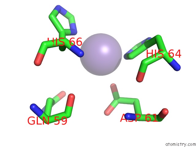
Mono view
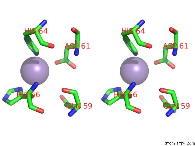
Stereo pair view
Mono view
Stereo pair view
A full contact list of Manganese with other atoms in the Mn binding
site number 1 of Crystal Structure of the ISHP608 Transposase in Complex with Right End 31-Mer Dna within 5.0Å range:
|
Reference:
O.Barabas,
D.R.Ronning,
C.Guynet,
A.B.Hickman,
B.Ton-Hoang,
M.Chandler,
F.Dyda.
Mechanism of IS200/IS605 Family Dna Transposases: Activation and Transposon-Directed Target Site Selection. Cell(Cambridge,Mass.) V. 132 208 2008.
ISSN: ISSN 0092-8674
PubMed: 18243097
DOI: 10.1016/J.CELL.2007.12.029
Page generated: Sat Oct 5 15:16:40 2024
ISSN: ISSN 0092-8674
PubMed: 18243097
DOI: 10.1016/J.CELL.2007.12.029
Last articles
Zn in 9J0NZn in 9J0O
Zn in 9J0P
Zn in 9FJX
Zn in 9EKB
Zn in 9C0F
Zn in 9CAH
Zn in 9CH0
Zn in 9CH3
Zn in 9CH1