Manganese »
PDB 2qjc-2v3y »
2tep »
Manganese in PDB 2tep: Peanut Lectin Complexed with T-Antigenic Disaccharide
Protein crystallography data
The structure of Peanut Lectin Complexed with T-Antigenic Disaccharide, PDB code: 2tep
was solved by
R.Ravishankar,
M.Ravindran,
K.Suguna,
A.Surolia,
M.Vijayan,
with X-Ray Crystallography technique. A brief refinement statistics is given in the table below:
| Resolution Low / High (Å) | 10.00 / 2.50 |
| Space group | P 21 21 2 |
| Cell size a, b, c (Å), α, β, γ (°) | 129.892, 126.676, 76.516, 90.00, 90.00, 90.00 |
| R / Rfree (%) | 17.5 / 25.1 |
Other elements in 2tep:
The structure of Peanut Lectin Complexed with T-Antigenic Disaccharide also contains other interesting chemical elements:
| Calcium | (Ca) | 4 atoms |
Manganese Binding Sites:
The binding sites of Manganese atom in the Peanut Lectin Complexed with T-Antigenic Disaccharide
(pdb code 2tep). This binding sites where shown within
5.0 Angstroms radius around Manganese atom.
In total 4 binding sites of Manganese where determined in the Peanut Lectin Complexed with T-Antigenic Disaccharide, PDB code: 2tep:
Jump to Manganese binding site number: 1; 2; 3; 4;
In total 4 binding sites of Manganese where determined in the Peanut Lectin Complexed with T-Antigenic Disaccharide, PDB code: 2tep:
Jump to Manganese binding site number: 1; 2; 3; 4;
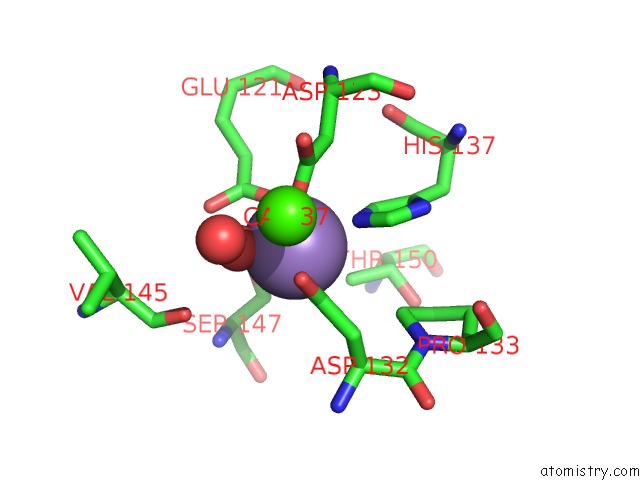
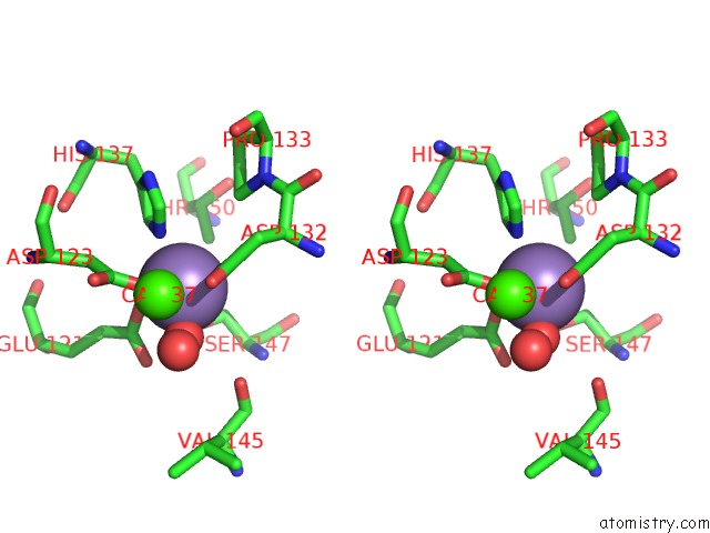
Manganese binding site 1 out of 4 in 2tep
Go back to
Manganese binding site 1 out
of 4 in the Peanut Lectin Complexed with T-Antigenic Disaccharide
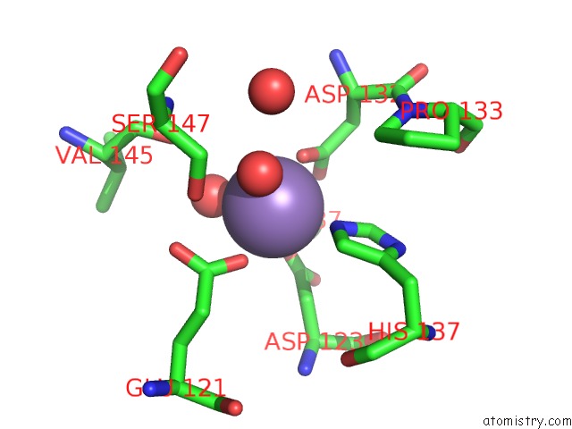
Mono view
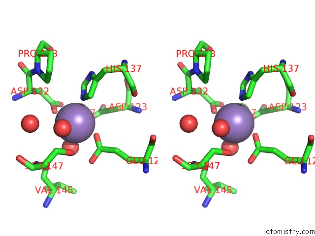
Stereo pair view
Mono view
Stereo pair view
A full contact list of Manganese with other atoms in the Mn binding
site number 1 of Peanut Lectin Complexed with T-Antigenic Disaccharide within 5.0Å range:
|
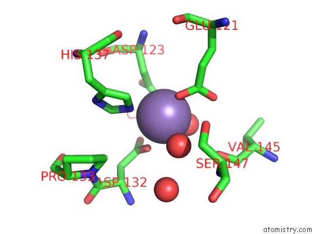
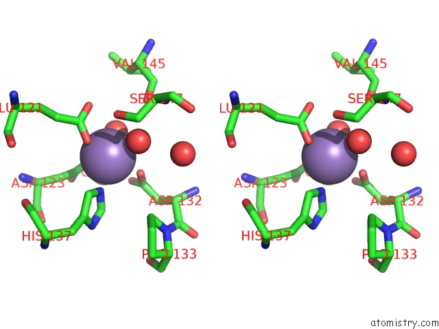
Manganese binding site 2 out of 4 in 2tep
Go back to
Manganese binding site 2 out
of 4 in the Peanut Lectin Complexed with T-Antigenic Disaccharide
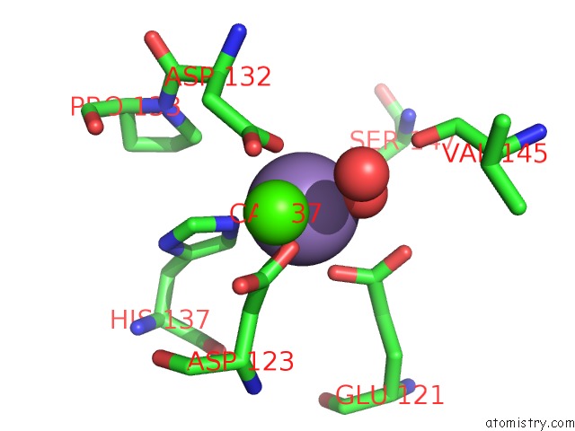
Mono view
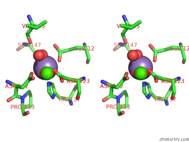
Stereo pair view
Mono view
Stereo pair view
A full contact list of Manganese with other atoms in the Mn binding
site number 2 of Peanut Lectin Complexed with T-Antigenic Disaccharide within 5.0Å range:
|
Manganese binding site 3 out of 4 in 2tep
Go back to
Manganese binding site 3 out
of 4 in the Peanut Lectin Complexed with T-Antigenic Disaccharide
Mono view
Stereo pair view
Mono view
Stereo pair view
A full contact list of Manganese with other atoms in the Mn binding
site number 3 of Peanut Lectin Complexed with T-Antigenic Disaccharide within 5.0Å range:
|
Manganese binding site 4 out of 4 in 2tep
Go back to
Manganese binding site 4 out
of 4 in the Peanut Lectin Complexed with T-Antigenic Disaccharide
Mono view
Stereo pair view
Mono view
Stereo pair view
A full contact list of Manganese with other atoms in the Mn binding
site number 4 of Peanut Lectin Complexed with T-Antigenic Disaccharide within 5.0Å range:
|
Reference:
R.Ravishankar,
M.Ravindran,
K.Suguna,
A.Surolia,
M.Vijayan.
The Specificity of Peanut Agglutinin For Thomsen-Friedenreich Antigen Is Mediated By Water-Bridges Curr.Sci. V. 72 855 1997.
ISSN: ISSN 0011-3891
Page generated: Sat Oct 5 15:11:03 2024
ISSN: ISSN 0011-3891
Last articles
Cl in 7UBRCl in 7UBJ
Cl in 7UBK
Cl in 7U9V
Cl in 7UAH
Cl in 7U98
Cl in 7U9F
Cl in 7U6H
Cl in 7U9A
Cl in 7U99