Manganese »
PDB 2qjc-2v3y »
2rcv »
Manganese in PDB 2rcv: Crystal Structure of the Bacillus Subtilis Superoxide Dismutase
Enzymatic activity of Crystal Structure of the Bacillus Subtilis Superoxide Dismutase
All present enzymatic activity of Crystal Structure of the Bacillus Subtilis Superoxide Dismutase:
1.15.1.1;
1.15.1.1;
Protein crystallography data
The structure of Crystal Structure of the Bacillus Subtilis Superoxide Dismutase, PDB code: 2rcv
was solved by
P.Liu,
H.E.Ewis,
Y.J.Huang,
C.D.Lu,
P.C.Tai,
I.T.Weber,
with X-Ray Crystallography technique. A brief refinement statistics is given in the table below:
| Resolution Low / High (Å) | 45.90 / 1.60 |
| Space group | P 1 |
| Cell size a, b, c (Å), α, β, γ (°) | 68.369, 84.034, 91.951, 99.13, 105.98, 105.58 |
| R / Rfree (%) | 21.1 / 23 |
Manganese Binding Sites:
The binding sites of Manganese atom in the Crystal Structure of the Bacillus Subtilis Superoxide Dismutase
(pdb code 2rcv). This binding sites where shown within
5.0 Angstroms radius around Manganese atom.
In total 8 binding sites of Manganese where determined in the Crystal Structure of the Bacillus Subtilis Superoxide Dismutase, PDB code: 2rcv:
Jump to Manganese binding site number: 1; 2; 3; 4; 5; 6; 7; 8;
In total 8 binding sites of Manganese where determined in the Crystal Structure of the Bacillus Subtilis Superoxide Dismutase, PDB code: 2rcv:
Jump to Manganese binding site number: 1; 2; 3; 4; 5; 6; 7; 8;
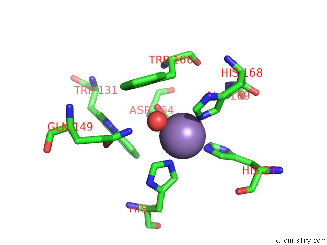
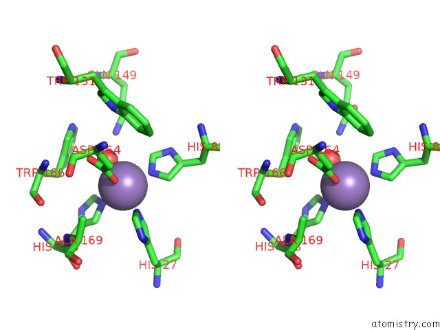
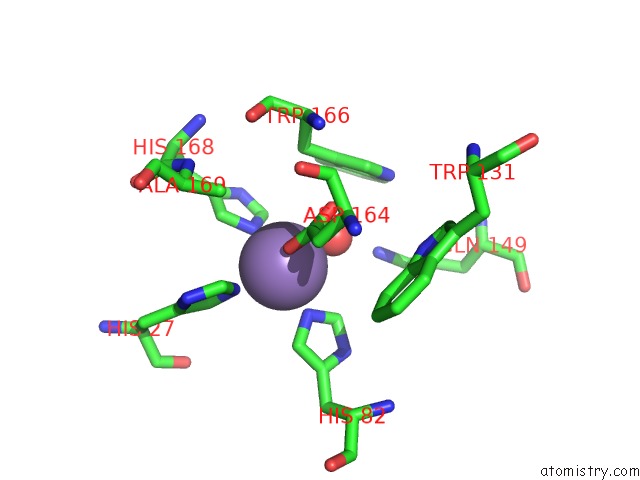
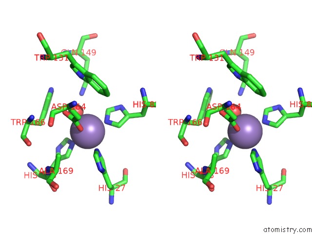
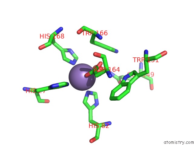
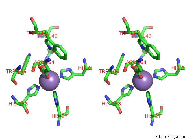
Manganese binding site 1 out of 8 in 2rcv
Go back to
Manganese binding site 1 out
of 8 in the Crystal Structure of the Bacillus Subtilis Superoxide Dismutase

Mono view
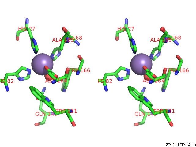
Stereo pair view
Mono view
Stereo pair view
A full contact list of Manganese with other atoms in the Mn binding
site number 1 of Crystal Structure of the Bacillus Subtilis Superoxide Dismutase within 5.0Å range:
|
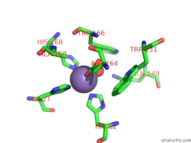
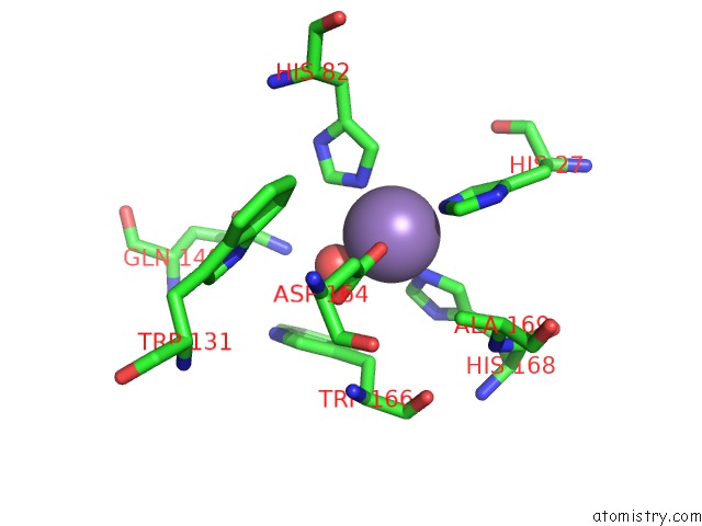
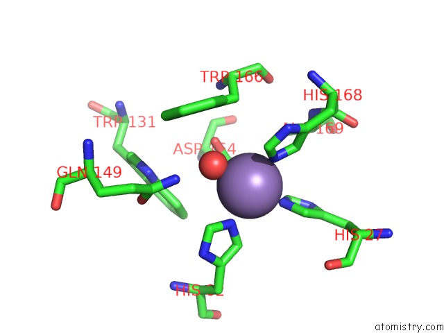
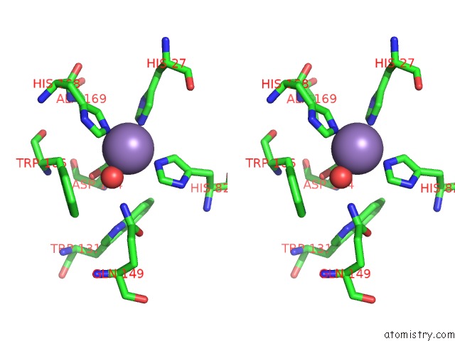
Manganese binding site 2 out of 8 in 2rcv
Go back to
Manganese binding site 2 out
of 8 in the Crystal Structure of the Bacillus Subtilis Superoxide Dismutase
Mono view
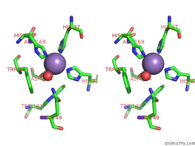
Stereo pair view
Mono view
Stereo pair view
A full contact list of Manganese with other atoms in the Mn binding
site number 2 of Crystal Structure of the Bacillus Subtilis Superoxide Dismutase within 5.0Å range:
|
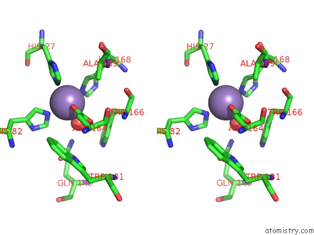
Manganese binding site 3 out of 8 in 2rcv
Go back to
Manganese binding site 3 out
of 8 in the Crystal Structure of the Bacillus Subtilis Superoxide Dismutase
Mono view
Stereo pair view
Mono view
Stereo pair view
A full contact list of Manganese with other atoms in the Mn binding
site number 3 of Crystal Structure of the Bacillus Subtilis Superoxide Dismutase within 5.0Å range:
|
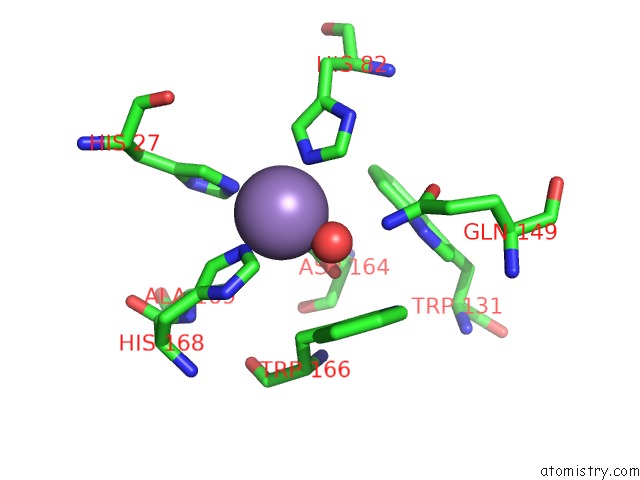
Manganese binding site 4 out of 8 in 2rcv
Go back to
Manganese binding site 4 out
of 8 in the Crystal Structure of the Bacillus Subtilis Superoxide Dismutase
Mono view
Stereo pair view
Mono view
Stereo pair view
A full contact list of Manganese with other atoms in the Mn binding
site number 4 of Crystal Structure of the Bacillus Subtilis Superoxide Dismutase within 5.0Å range:
|
Manganese binding site 5 out of 8 in 2rcv
Go back to
Manganese binding site 5 out
of 8 in the Crystal Structure of the Bacillus Subtilis Superoxide Dismutase
Mono view

Stereo pair view
Mono view
Stereo pair view
A full contact list of Manganese with other atoms in the Mn binding
site number 5 of Crystal Structure of the Bacillus Subtilis Superoxide Dismutase within 5.0Å range:
|
Manganese binding site 6 out of 8 in 2rcv
Go back to
Manganese binding site 6 out
of 8 in the Crystal Structure of the Bacillus Subtilis Superoxide Dismutase
Mono view
Stereo pair view
Mono view
Stereo pair view
A full contact list of Manganese with other atoms in the Mn binding
site number 6 of Crystal Structure of the Bacillus Subtilis Superoxide Dismutase within 5.0Å range:
|
Manganese binding site 7 out of 8 in 2rcv
Go back to
Manganese binding site 7 out
of 8 in the Crystal Structure of the Bacillus Subtilis Superoxide Dismutase
Mono view
Stereo pair view
Mono view
Stereo pair view
A full contact list of Manganese with other atoms in the Mn binding
site number 7 of Crystal Structure of the Bacillus Subtilis Superoxide Dismutase within 5.0Å range:
|
Manganese binding site 8 out of 8 in 2rcv
Go back to
Manganese binding site 8 out
of 8 in the Crystal Structure of the Bacillus Subtilis Superoxide Dismutase
Mono view
Stereo pair view
Mono view
Stereo pair view
A full contact list of Manganese with other atoms in the Mn binding
site number 8 of Crystal Structure of the Bacillus Subtilis Superoxide Dismutase within 5.0Å range:
|
Reference:
P.Liu,
H.E.Ewis,
Y.J.Huang,
C.D.Lu,
P.C.Tai,
I.T.Weber.
Structure of Bacillus Subtilis Superoxide Dismutase. Acta Crystallogr.,Sect.F V. 63 1003 2007.
ISSN: ESSN 1744-3091
PubMed: 18084079
DOI: 10.1107/S1744309107054127
Page generated: Sat Oct 5 15:07:48 2024
ISSN: ESSN 1744-3091
PubMed: 18084079
DOI: 10.1107/S1744309107054127
Last articles
Fe in 2YXOFe in 2YRS
Fe in 2YXC
Fe in 2YNM
Fe in 2YVJ
Fe in 2YP1
Fe in 2YU2
Fe in 2YU1
Fe in 2YQB
Fe in 2YOO