Manganese »
PDB 2qjc-2v3y »
2qum »
Manganese in PDB 2qum: Crystal Structure of D-Tagatose 3-Epimerase From Pseudomonas Cichorii with D-Tagatose
Protein crystallography data
The structure of Crystal Structure of D-Tagatose 3-Epimerase From Pseudomonas Cichorii with D-Tagatose, PDB code: 2qum
was solved by
H.Yoshida,
M.Yamada,
T.Nishitani,
G.Takada,
K.Izumori,
S.Kamitori,
with X-Ray Crystallography technique. A brief refinement statistics is given in the table below:
| Resolution Low / High (Å) | 41.31 / 2.28 |
| Space group | P 1 21 1 |
| Cell size a, b, c (Å), α, β, γ (°) | 49.414, 139.432, 92.860, 90.00, 104.04, 90.00 |
| R / Rfree (%) | 18.5 / 23.9 |
Manganese Binding Sites:
The binding sites of Manganese atom in the Crystal Structure of D-Tagatose 3-Epimerase From Pseudomonas Cichorii with D-Tagatose
(pdb code 2qum). This binding sites where shown within
5.0 Angstroms radius around Manganese atom.
In total 4 binding sites of Manganese where determined in the Crystal Structure of D-Tagatose 3-Epimerase From Pseudomonas Cichorii with D-Tagatose, PDB code: 2qum:
Jump to Manganese binding site number: 1; 2; 3; 4;
In total 4 binding sites of Manganese where determined in the Crystal Structure of D-Tagatose 3-Epimerase From Pseudomonas Cichorii with D-Tagatose, PDB code: 2qum:
Jump to Manganese binding site number: 1; 2; 3; 4;
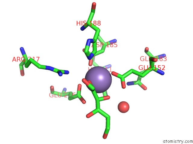
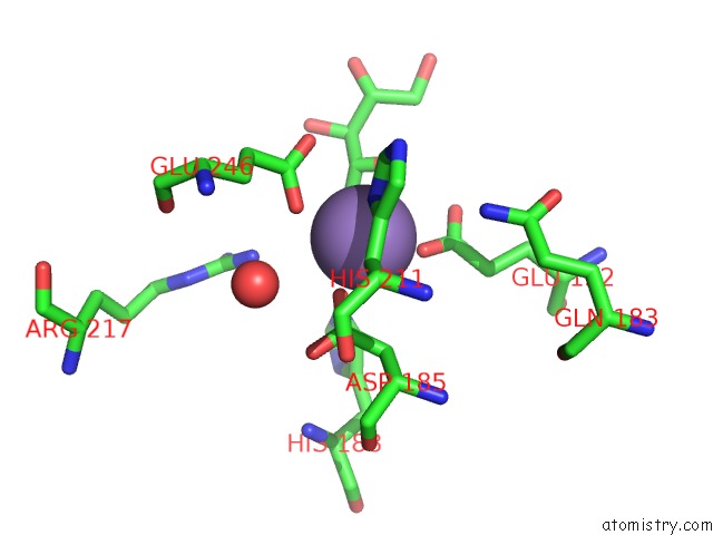
Manganese binding site 1 out of 4 in 2qum
Go back to
Manganese binding site 1 out
of 4 in the Crystal Structure of D-Tagatose 3-Epimerase From Pseudomonas Cichorii with D-Tagatose
Mono view
Stereo pair view
Mono view
Stereo pair view
A full contact list of Manganese with other atoms in the Mn binding
site number 1 of Crystal Structure of D-Tagatose 3-Epimerase From Pseudomonas Cichorii with D-Tagatose within 5.0Å range:
|
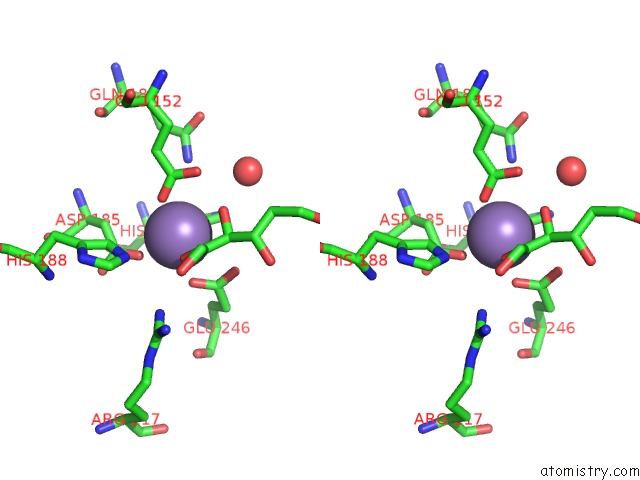
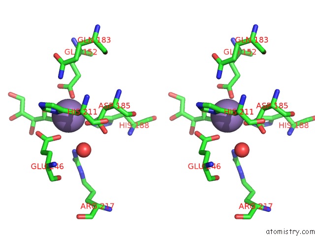
Manganese binding site 2 out of 4 in 2qum
Go back to
Manganese binding site 2 out
of 4 in the Crystal Structure of D-Tagatose 3-Epimerase From Pseudomonas Cichorii with D-Tagatose
Mono view
Stereo pair view
Mono view
Stereo pair view
A full contact list of Manganese with other atoms in the Mn binding
site number 2 of Crystal Structure of D-Tagatose 3-Epimerase From Pseudomonas Cichorii with D-Tagatose within 5.0Å range:
|
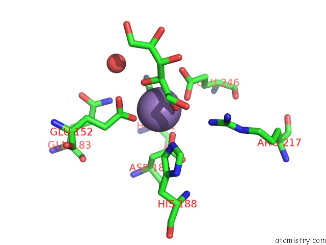
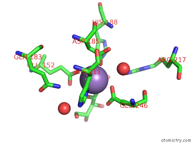
Manganese binding site 3 out of 4 in 2qum
Go back to
Manganese binding site 3 out
of 4 in the Crystal Structure of D-Tagatose 3-Epimerase From Pseudomonas Cichorii with D-Tagatose
Mono view
Stereo pair view
Mono view
Stereo pair view
A full contact list of Manganese with other atoms in the Mn binding
site number 3 of Crystal Structure of D-Tagatose 3-Epimerase From Pseudomonas Cichorii with D-Tagatose within 5.0Å range:
|
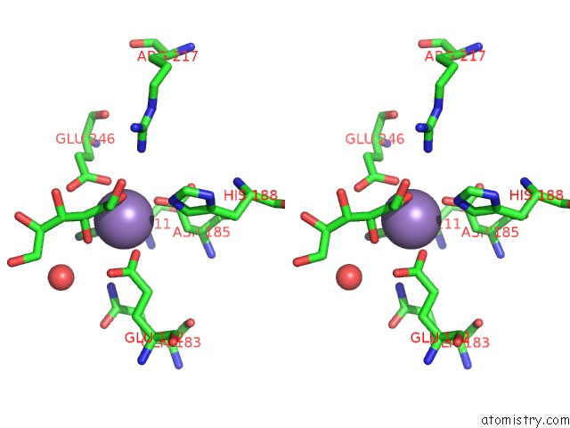
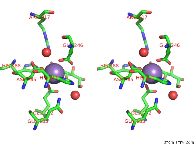
Manganese binding site 4 out of 4 in 2qum
Go back to
Manganese binding site 4 out
of 4 in the Crystal Structure of D-Tagatose 3-Epimerase From Pseudomonas Cichorii with D-Tagatose
Mono view
Stereo pair view
Mono view
Stereo pair view
A full contact list of Manganese with other atoms in the Mn binding
site number 4 of Crystal Structure of D-Tagatose 3-Epimerase From Pseudomonas Cichorii with D-Tagatose within 5.0Å range:
|
Reference:
H.Yoshida,
M.Yamada,
T.Nishitani,
G.Takada,
K.Izumori,
S.Kamitori.
Crystal Structures of D-Tagatose 3-Epimerase From Pseudomonas Cichorii and Its Complexes with D-Tagatose and D-Fructose J.Mol.Biol. V. 374 443 2007.
ISSN: ISSN 0022-2836
PubMed: 17936787
DOI: 10.1016/J.JMB.2007.09.033
Page generated: Sat Oct 5 15:05:31 2024
ISSN: ISSN 0022-2836
PubMed: 17936787
DOI: 10.1016/J.JMB.2007.09.033
Last articles
Zn in 9JYWZn in 9IR4
Zn in 9IR3
Zn in 9GMX
Zn in 9GMW
Zn in 9JEJ
Zn in 9ERF
Zn in 9ERE
Zn in 9EGV
Zn in 9EGW