Manganese »
PDB 2pal-2qgi »
2qcs »
Manganese in PDB 2qcs: A Complex Structure Between the Catalytic and Regulatory Subunit of Protein Kinase A That Represents the Inhibited State
Enzymatic activity of A Complex Structure Between the Catalytic and Regulatory Subunit of Protein Kinase A That Represents the Inhibited State
All present enzymatic activity of A Complex Structure Between the Catalytic and Regulatory Subunit of Protein Kinase A That Represents the Inhibited State:
2.7.11.11;
2.7.11.11;
Protein crystallography data
The structure of A Complex Structure Between the Catalytic and Regulatory Subunit of Protein Kinase A That Represents the Inhibited State, PDB code: 2qcs
was solved by
C.Kim,
C.Y.Cheng,
A.S.Saldanha,
S.S.Taylor,
with X-Ray Crystallography technique. A brief refinement statistics is given in the table below:
| Resolution Low / High (Å) | 50.00 / 2.20 |
| Space group | P 32 2 1 |
| Cell size a, b, c (Å), α, β, γ (°) | 125.809, 125.809, 140.941, 90.00, 90.00, 120.00 |
| R / Rfree (%) | 19.2 / 22.5 |
Manganese Binding Sites:
The binding sites of Manganese atom in the A Complex Structure Between the Catalytic and Regulatory Subunit of Protein Kinase A That Represents the Inhibited State
(pdb code 2qcs). This binding sites where shown within
5.0 Angstroms radius around Manganese atom.
In total 2 binding sites of Manganese where determined in the A Complex Structure Between the Catalytic and Regulatory Subunit of Protein Kinase A That Represents the Inhibited State, PDB code: 2qcs:
Jump to Manganese binding site number: 1; 2;
In total 2 binding sites of Manganese where determined in the A Complex Structure Between the Catalytic and Regulatory Subunit of Protein Kinase A That Represents the Inhibited State, PDB code: 2qcs:
Jump to Manganese binding site number: 1; 2;
Manganese binding site 1 out of 2 in 2qcs
Go back to
Manganese binding site 1 out
of 2 in the A Complex Structure Between the Catalytic and Regulatory Subunit of Protein Kinase A That Represents the Inhibited State
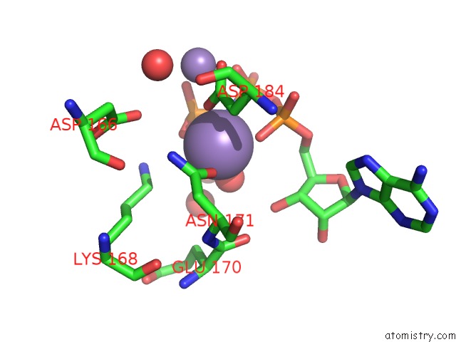
Mono view
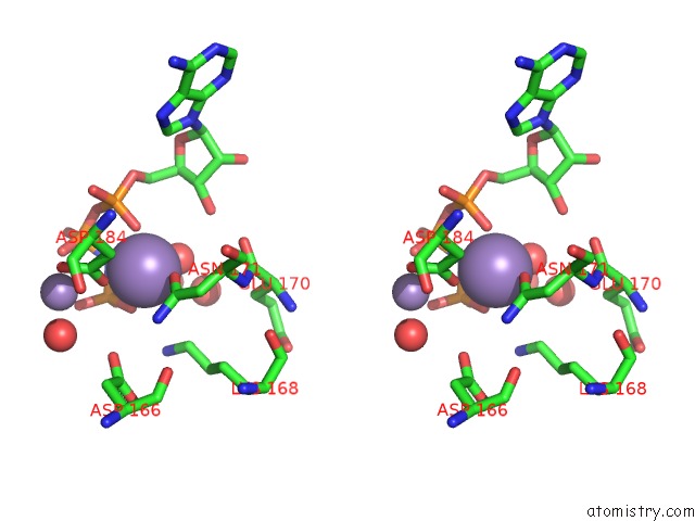
Stereo pair view
Mono view
Stereo pair view
A full contact list of Manganese with other atoms in the Mn binding
site number 1 of A Complex Structure Between the Catalytic and Regulatory Subunit of Protein Kinase A That Represents the Inhibited State within 5.0Å range:
|
Manganese binding site 2 out of 2 in 2qcs
Go back to
Manganese binding site 2 out
of 2 in the A Complex Structure Between the Catalytic and Regulatory Subunit of Protein Kinase A That Represents the Inhibited State
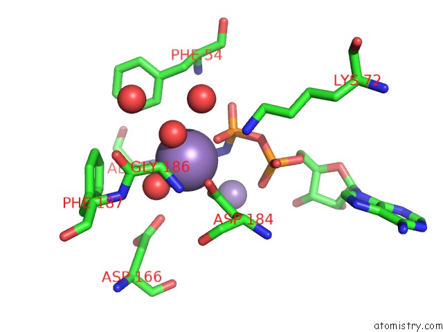
Mono view
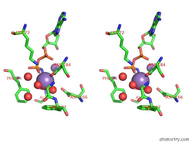
Stereo pair view
Mono view
Stereo pair view
A full contact list of Manganese with other atoms in the Mn binding
site number 2 of A Complex Structure Between the Catalytic and Regulatory Subunit of Protein Kinase A That Represents the Inhibited State within 5.0Å range:
|
Reference:
C.Kim,
C.Y.Cheng,
S.A.Saldanha,
S.S.Taylor.
Pka-I Holoenzyme Structure Reveals A Mechanism For Camp-Dependent Activation. Cell(Cambridge,Mass.) V. 130 1032 2007.
ISSN: ISSN 0092-8674
PubMed: 17889648
DOI: 10.1016/J.CELL.2007.07.018
Page generated: Sat Oct 5 14:59:55 2024
ISSN: ISSN 0092-8674
PubMed: 17889648
DOI: 10.1016/J.CELL.2007.07.018
Last articles
Zn in 9J0NZn in 9J0O
Zn in 9J0P
Zn in 9FJX
Zn in 9EKB
Zn in 9C0F
Zn in 9CAH
Zn in 9CH0
Zn in 9CH3
Zn in 9CH1