Manganese »
PDB 2pal-2qgi »
2pyj »
Manganese in PDB 2pyj: PHI29 Dna Polymerase Complexed with Primer-Template Dna and Incoming Nucleotide Substrates (Ternary Complex)
Enzymatic activity of PHI29 Dna Polymerase Complexed with Primer-Template Dna and Incoming Nucleotide Substrates (Ternary Complex)
All present enzymatic activity of PHI29 Dna Polymerase Complexed with Primer-Template Dna and Incoming Nucleotide Substrates (Ternary Complex):
2.7.7.7;
2.7.7.7;
Protein crystallography data
The structure of PHI29 Dna Polymerase Complexed with Primer-Template Dna and Incoming Nucleotide Substrates (Ternary Complex), PDB code: 2pyj
was solved by
A.J.Berman,
S.Kamtekar,
J.L.Goodman,
J.M.Lazaro,
M.De Vega,
L.Blanco,
M.Salas,
T.A.Steitz,
with X-Ray Crystallography technique. A brief refinement statistics is given in the table below:
| Resolution Low / High (Å) | 41.07 / 2.03 |
| Space group | P 1 21 1 |
| Cell size a, b, c (Å), α, β, γ (°) | 72.835, 114.667, 104.761, 90.00, 94.07, 90.00 |
| R / Rfree (%) | 18.9 / 23.4 |
Other elements in 2pyj:
The structure of PHI29 Dna Polymerase Complexed with Primer-Template Dna and Incoming Nucleotide Substrates (Ternary Complex) also contains other interesting chemical elements:
| Magnesium | (Mg) | 2 atoms |
Manganese Binding Sites:
The binding sites of Manganese atom in the PHI29 Dna Polymerase Complexed with Primer-Template Dna and Incoming Nucleotide Substrates (Ternary Complex)
(pdb code 2pyj). This binding sites where shown within
5.0 Angstroms radius around Manganese atom.
In total 2 binding sites of Manganese where determined in the PHI29 Dna Polymerase Complexed with Primer-Template Dna and Incoming Nucleotide Substrates (Ternary Complex), PDB code: 2pyj:
Jump to Manganese binding site number: 1; 2;
In total 2 binding sites of Manganese where determined in the PHI29 Dna Polymerase Complexed with Primer-Template Dna and Incoming Nucleotide Substrates (Ternary Complex), PDB code: 2pyj:
Jump to Manganese binding site number: 1; 2;
Manganese binding site 1 out of 2 in 2pyj
Go back to
Manganese binding site 1 out
of 2 in the PHI29 Dna Polymerase Complexed with Primer-Template Dna and Incoming Nucleotide Substrates (Ternary Complex)
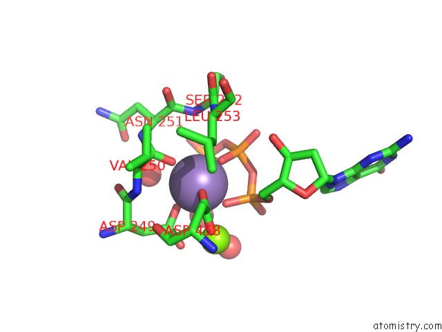
Mono view
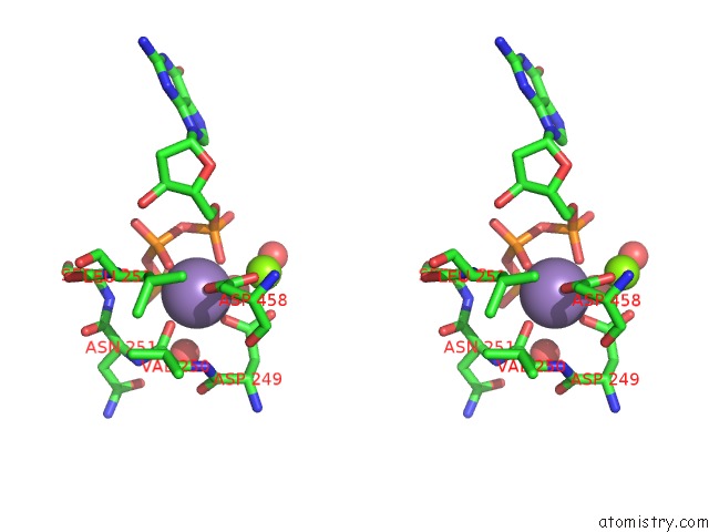
Stereo pair view
Mono view
Stereo pair view
A full contact list of Manganese with other atoms in the Mn binding
site number 1 of PHI29 Dna Polymerase Complexed with Primer-Template Dna and Incoming Nucleotide Substrates (Ternary Complex) within 5.0Å range:
|
Manganese binding site 2 out of 2 in 2pyj
Go back to
Manganese binding site 2 out
of 2 in the PHI29 Dna Polymerase Complexed with Primer-Template Dna and Incoming Nucleotide Substrates (Ternary Complex)
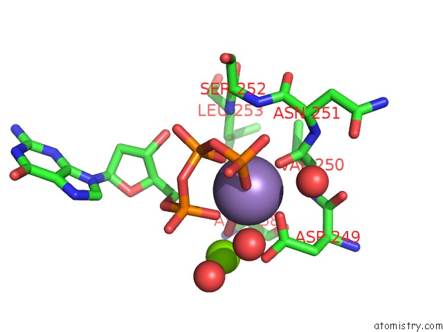
Mono view
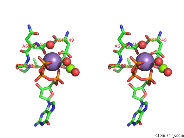
Stereo pair view
Mono view
Stereo pair view
A full contact list of Manganese with other atoms in the Mn binding
site number 2 of PHI29 Dna Polymerase Complexed with Primer-Template Dna and Incoming Nucleotide Substrates (Ternary Complex) within 5.0Å range:
|
Reference:
A.J.Berman,
S.Kamtekar,
J.L.Goodman,
J.M.Lazaro,
M.De Vega,
L.Blanco,
M.Salas,
T.A.Steitz.
Structures of PHI29 Dna Polymerase Complexed with Substrate: the Mechanism of Translocation in B-Family Polymerases Embo J. V. 26 3494 2007.
ISSN: ISSN 0261-4189
PubMed: 17611604
DOI: 10.1038/SJ.EMBOJ.7601780
Page generated: Sat Oct 5 14:57:47 2024
ISSN: ISSN 0261-4189
PubMed: 17611604
DOI: 10.1038/SJ.EMBOJ.7601780
Last articles
Ca in 5MQMCa in 5MQN
Ca in 5MPY
Ca in 5MOR
Ca in 5MOS
Ca in 5MOQ
Ca in 5MOO
Ca in 5MOP
Ca in 5MNR
Ca in 5MON