Manganese »
PDB 2pal-2qgi »
2pho »
Manganese in PDB 2pho: Crystal Structure of Human Arginase I Complexed with Thiosemicarbazide at 1.95 Resolution
Enzymatic activity of Crystal Structure of Human Arginase I Complexed with Thiosemicarbazide at 1.95 Resolution
All present enzymatic activity of Crystal Structure of Human Arginase I Complexed with Thiosemicarbazide at 1.95 Resolution:
3.5.3.1;
3.5.3.1;
Protein crystallography data
The structure of Crystal Structure of Human Arginase I Complexed with Thiosemicarbazide at 1.95 Resolution, PDB code: 2pho
was solved by
L.Di Costanzo,
D.W.Christianson,
with X-Ray Crystallography technique. A brief refinement statistics is given in the table below:
| Resolution Low / High (Å) | 34.18 / 1.95 |
| Space group | P 3 |
| Cell size a, b, c (Å), α, β, γ (°) | 90.610, 90.610, 69.630, 90.00, 90.00, 120.00 |
| R / Rfree (%) | 16.9 / 21.9 |
Manganese Binding Sites:
The binding sites of Manganese atom in the Crystal Structure of Human Arginase I Complexed with Thiosemicarbazide at 1.95 Resolution
(pdb code 2pho). This binding sites where shown within
5.0 Angstroms radius around Manganese atom.
In total 4 binding sites of Manganese where determined in the Crystal Structure of Human Arginase I Complexed with Thiosemicarbazide at 1.95 Resolution, PDB code: 2pho:
Jump to Manganese binding site number: 1; 2; 3; 4;
In total 4 binding sites of Manganese where determined in the Crystal Structure of Human Arginase I Complexed with Thiosemicarbazide at 1.95 Resolution, PDB code: 2pho:
Jump to Manganese binding site number: 1; 2; 3; 4;
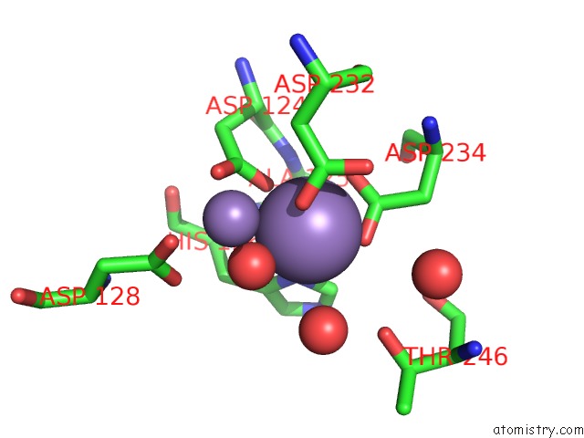
Manganese binding site 1 out of 4 in 2pho
Go back to
Manganese binding site 1 out
of 4 in the Crystal Structure of Human Arginase I Complexed with Thiosemicarbazide at 1.95 Resolution
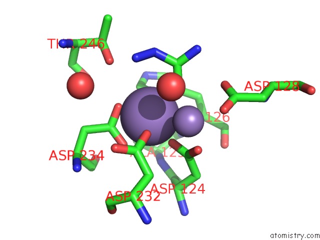
Mono view
Stereo pair view
Mono view
Stereo pair view
A full contact list of Manganese with other atoms in the Mn binding
site number 1 of Crystal Structure of Human Arginase I Complexed with Thiosemicarbazide at 1.95 Resolution within 5.0Å range:
|
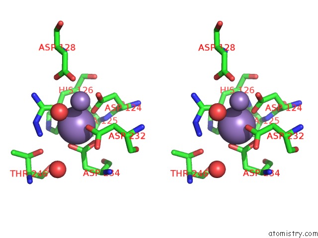
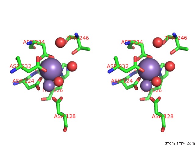
Manganese binding site 2 out of 4 in 2pho
Go back to
Manganese binding site 2 out
of 4 in the Crystal Structure of Human Arginase I Complexed with Thiosemicarbazide at 1.95 Resolution
Mono view
Stereo pair view
Mono view
Stereo pair view
A full contact list of Manganese with other atoms in the Mn binding
site number 2 of Crystal Structure of Human Arginase I Complexed with Thiosemicarbazide at 1.95 Resolution within 5.0Å range:
|
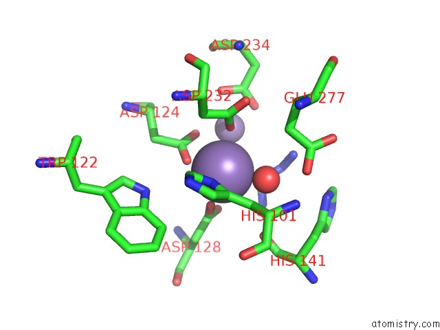
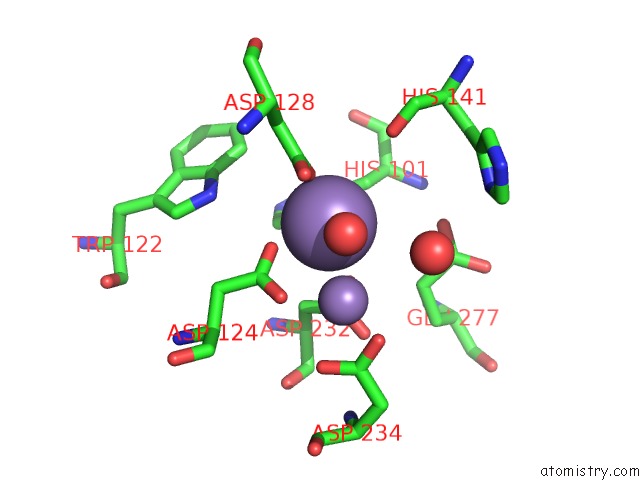
Manganese binding site 3 out of 4 in 2pho
Go back to
Manganese binding site 3 out
of 4 in the Crystal Structure of Human Arginase I Complexed with Thiosemicarbazide at 1.95 Resolution
Mono view
Stereo pair view
Mono view
Stereo pair view
A full contact list of Manganese with other atoms in the Mn binding
site number 3 of Crystal Structure of Human Arginase I Complexed with Thiosemicarbazide at 1.95 Resolution within 5.0Å range:
|
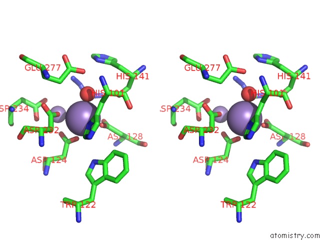
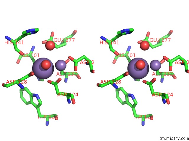
Manganese binding site 4 out of 4 in 2pho
Go back to
Manganese binding site 4 out
of 4 in the Crystal Structure of Human Arginase I Complexed with Thiosemicarbazide at 1.95 Resolution
Mono view
Stereo pair view
Mono view
Stereo pair view
A full contact list of Manganese with other atoms in the Mn binding
site number 4 of Crystal Structure of Human Arginase I Complexed with Thiosemicarbazide at 1.95 Resolution within 5.0Å range:
|
Reference:
L.Di Costanzo,
M.E.Pique,
D.W.Christianson.
Crystal Structure of Human Arginase I Complexed with Thiosemicarbazide Reveals An Unusual Thiocarbonyl Mu-Sulfide Ligand in the Binuclear Manganese Cluster. J.Am.Chem.Soc. V. 129 6388 2007.
ISSN: ISSN 0002-7863
PubMed: 17469833
DOI: 10.1021/JA071567J
Page generated: Sat Oct 5 14:54:42 2024
ISSN: ISSN 0002-7863
PubMed: 17469833
DOI: 10.1021/JA071567J
Last articles
Zn in 9J0NZn in 9J0O
Zn in 9J0P
Zn in 9FJX
Zn in 9EKB
Zn in 9C0F
Zn in 9CAH
Zn in 9CH0
Zn in 9CH3
Zn in 9CH1