Manganese »
PDB 2pal-2qgi »
2phk »
Manganese in PDB 2phk: The Crystal Structure of A Phosphorylase Kinase Peptide Substrate Complex: Kinase Substrate Recognition
Enzymatic activity of The Crystal Structure of A Phosphorylase Kinase Peptide Substrate Complex: Kinase Substrate Recognition
All present enzymatic activity of The Crystal Structure of A Phosphorylase Kinase Peptide Substrate Complex: Kinase Substrate Recognition:
2.7.1.38;
2.7.1.38;
Protein crystallography data
The structure of The Crystal Structure of A Phosphorylase Kinase Peptide Substrate Complex: Kinase Substrate Recognition, PDB code: 2phk
was solved by
E.D.Lowe,
M.E.M.Noble,
V.T.Skamnaki,
N.G.Oikonomakos,
D.J.Owen,
L.N.Johnson,
with X-Ray Crystallography technique. A brief refinement statistics is given in the table below:
| Resolution Low / High (Å) | 25.00 / 2.60 |
| Space group | P 32 2 1 |
| Cell size a, b, c (Å), α, β, γ (°) | 65.300, 65.300, 145.800, 90.00, 90.00, 120.00 |
| R / Rfree (%) | 23.6 / 30 |
Manganese Binding Sites:
The binding sites of Manganese atom in the The Crystal Structure of A Phosphorylase Kinase Peptide Substrate Complex: Kinase Substrate Recognition
(pdb code 2phk). This binding sites where shown within
5.0 Angstroms radius around Manganese atom.
In total 2 binding sites of Manganese where determined in the The Crystal Structure of A Phosphorylase Kinase Peptide Substrate Complex: Kinase Substrate Recognition, PDB code: 2phk:
Jump to Manganese binding site number: 1; 2;
In total 2 binding sites of Manganese where determined in the The Crystal Structure of A Phosphorylase Kinase Peptide Substrate Complex: Kinase Substrate Recognition, PDB code: 2phk:
Jump to Manganese binding site number: 1; 2;
Manganese binding site 1 out of 2 in 2phk
Go back to
Manganese binding site 1 out
of 2 in the The Crystal Structure of A Phosphorylase Kinase Peptide Substrate Complex: Kinase Substrate Recognition
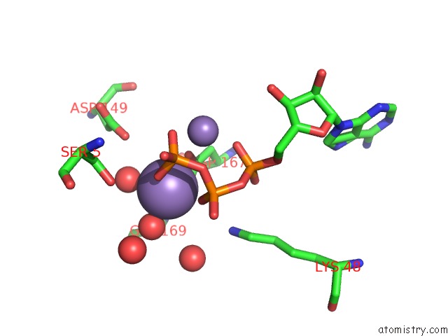
Mono view
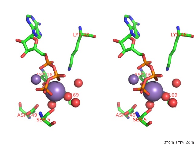
Stereo pair view
Mono view
Stereo pair view
A full contact list of Manganese with other atoms in the Mn binding
site number 1 of The Crystal Structure of A Phosphorylase Kinase Peptide Substrate Complex: Kinase Substrate Recognition within 5.0Å range:
|
Manganese binding site 2 out of 2 in 2phk
Go back to
Manganese binding site 2 out
of 2 in the The Crystal Structure of A Phosphorylase Kinase Peptide Substrate Complex: Kinase Substrate Recognition
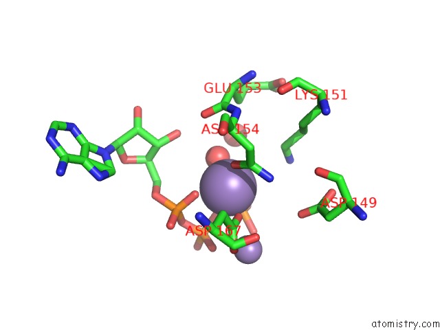
Mono view
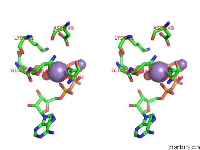
Stereo pair view
Mono view
Stereo pair view
A full contact list of Manganese with other atoms in the Mn binding
site number 2 of The Crystal Structure of A Phosphorylase Kinase Peptide Substrate Complex: Kinase Substrate Recognition within 5.0Å range:
|
Reference:
E.D.Lowe,
M.E.Noble,
V.T.Skamnaki,
N.G.Oikonomakos,
D.J.Owen,
L.N.Johnson.
The Crystal Structure of A Phosphorylase Kinase Peptide Substrate Complex: Kinase Substrate Recognition. Embo J. V. 16 6646 1997.
ISSN: ISSN 0261-4189
PubMed: 9362479
DOI: 10.1093/EMBOJ/16.22.6646
Page generated: Sat Oct 5 14:53:54 2024
ISSN: ISSN 0261-4189
PubMed: 9362479
DOI: 10.1093/EMBOJ/16.22.6646
Last articles
Cl in 7TMUCl in 7TJP
Cl in 7TMZ
Cl in 7TLG
Cl in 7TJC
Cl in 7TJO
Cl in 7TLE
Cl in 7TKV
Cl in 7THH
Cl in 7TIV