Manganese »
PDB 2nzd-2p9a »
2p7p »
Manganese in PDB 2p7p: Crystal Structure of Genomically Encoded Fosfomycin Resistance Protein, Fosx, From Listeria Monocytogenes Complexed with Mn(II) and Sulfate Ion
Protein crystallography data
The structure of Crystal Structure of Genomically Encoded Fosfomycin Resistance Protein, Fosx, From Listeria Monocytogenes Complexed with Mn(II) and Sulfate Ion, PDB code: 2p7p
was solved by
K.L.Fillgrove,
S.Pakhomova,
M.Schaab,
M.E.Newcomer,
R.N.Armstrong,
with X-Ray Crystallography technique. A brief refinement statistics is given in the table below:
| Resolution Low / High (Å) | 23.08 / 2.17 |
| Space group | C 1 2 1 |
| Cell size a, b, c (Å), α, β, γ (°) | 169.500, 70.565, 84.145, 90.00, 113.74, 90.00 |
| R / Rfree (%) | 22.6 / 27 |
Manganese Binding Sites:
The binding sites of Manganese atom in the Crystal Structure of Genomically Encoded Fosfomycin Resistance Protein, Fosx, From Listeria Monocytogenes Complexed with Mn(II) and Sulfate Ion
(pdb code 2p7p). This binding sites where shown within
5.0 Angstroms radius around Manganese atom.
In total 6 binding sites of Manganese where determined in the Crystal Structure of Genomically Encoded Fosfomycin Resistance Protein, Fosx, From Listeria Monocytogenes Complexed with Mn(II) and Sulfate Ion, PDB code: 2p7p:
Jump to Manganese binding site number: 1; 2; 3; 4; 5; 6;
In total 6 binding sites of Manganese where determined in the Crystal Structure of Genomically Encoded Fosfomycin Resistance Protein, Fosx, From Listeria Monocytogenes Complexed with Mn(II) and Sulfate Ion, PDB code: 2p7p:
Jump to Manganese binding site number: 1; 2; 3; 4; 5; 6;
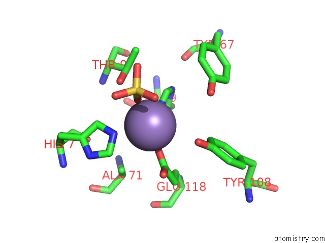
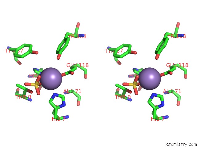
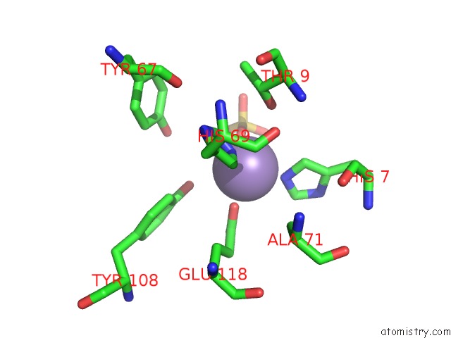
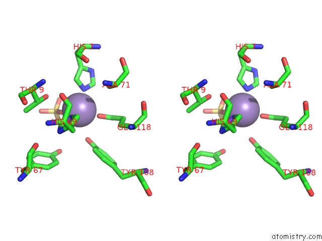
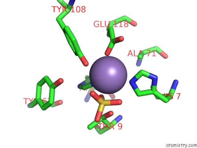
Manganese binding site 1 out of 6 in 2p7p
Go back to
Manganese binding site 1 out
of 6 in the Crystal Structure of Genomically Encoded Fosfomycin Resistance Protein, Fosx, From Listeria Monocytogenes Complexed with Mn(II) and Sulfate Ion
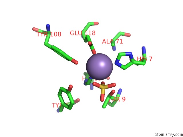
Mono view
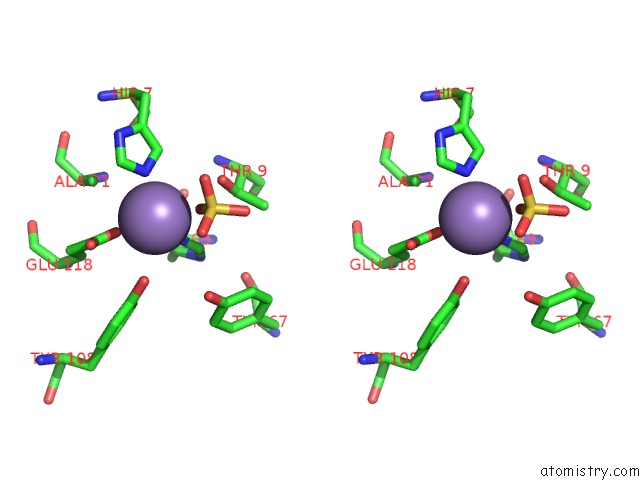
Stereo pair view
Mono view
Stereo pair view
A full contact list of Manganese with other atoms in the Mn binding
site number 1 of Crystal Structure of Genomically Encoded Fosfomycin Resistance Protein, Fosx, From Listeria Monocytogenes Complexed with Mn(II) and Sulfate Ion within 5.0Å range:
|
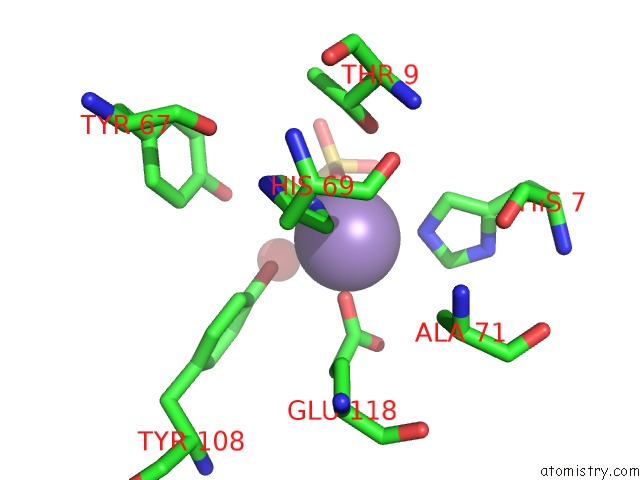
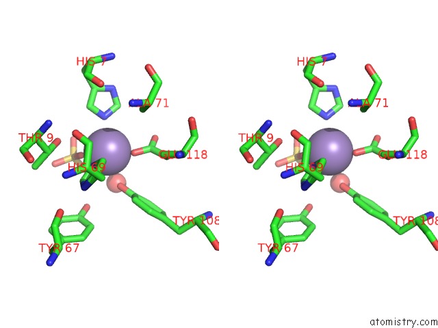
Manganese binding site 2 out of 6 in 2p7p
Go back to
Manganese binding site 2 out
of 6 in the Crystal Structure of Genomically Encoded Fosfomycin Resistance Protein, Fosx, From Listeria Monocytogenes Complexed with Mn(II) and Sulfate Ion
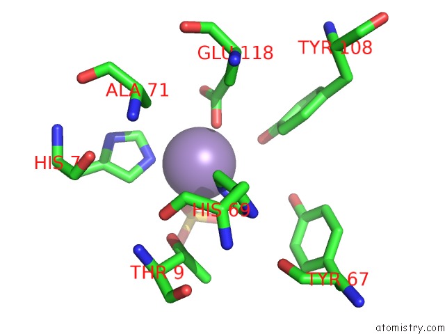
Mono view
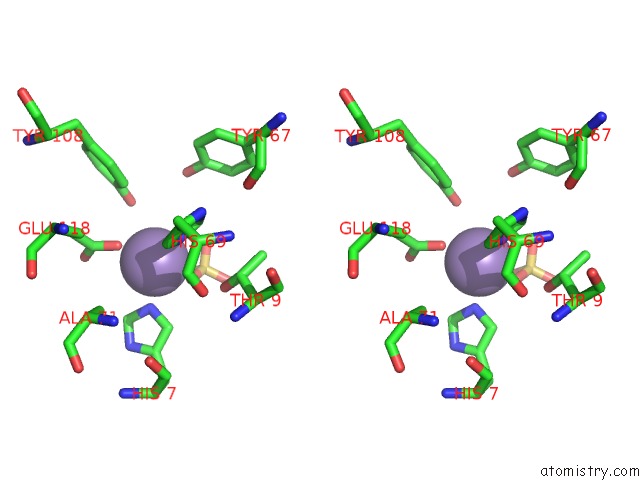
Stereo pair view
Mono view
Stereo pair view
A full contact list of Manganese with other atoms in the Mn binding
site number 2 of Crystal Structure of Genomically Encoded Fosfomycin Resistance Protein, Fosx, From Listeria Monocytogenes Complexed with Mn(II) and Sulfate Ion within 5.0Å range:
|
Manganese binding site 3 out of 6 in 2p7p
Go back to
Manganese binding site 3 out
of 6 in the Crystal Structure of Genomically Encoded Fosfomycin Resistance Protein, Fosx, From Listeria Monocytogenes Complexed with Mn(II) and Sulfate Ion
Mono view
Stereo pair view
Mono view
Stereo pair view
A full contact list of Manganese with other atoms in the Mn binding
site number 3 of Crystal Structure of Genomically Encoded Fosfomycin Resistance Protein, Fosx, From Listeria Monocytogenes Complexed with Mn(II) and Sulfate Ion within 5.0Å range:
|
Manganese binding site 4 out of 6 in 2p7p
Go back to
Manganese binding site 4 out
of 6 in the Crystal Structure of Genomically Encoded Fosfomycin Resistance Protein, Fosx, From Listeria Monocytogenes Complexed with Mn(II) and Sulfate Ion
Mono view
Stereo pair view
Mono view
Stereo pair view
A full contact list of Manganese with other atoms in the Mn binding
site number 4 of Crystal Structure of Genomically Encoded Fosfomycin Resistance Protein, Fosx, From Listeria Monocytogenes Complexed with Mn(II) and Sulfate Ion within 5.0Å range:
|
Manganese binding site 5 out of 6 in 2p7p
Go back to
Manganese binding site 5 out
of 6 in the Crystal Structure of Genomically Encoded Fosfomycin Resistance Protein, Fosx, From Listeria Monocytogenes Complexed with Mn(II) and Sulfate Ion
Mono view
Stereo pair view
Mono view
Stereo pair view
A full contact list of Manganese with other atoms in the Mn binding
site number 5 of Crystal Structure of Genomically Encoded Fosfomycin Resistance Protein, Fosx, From Listeria Monocytogenes Complexed with Mn(II) and Sulfate Ion within 5.0Å range:
|
Manganese binding site 6 out of 6 in 2p7p
Go back to
Manganese binding site 6 out
of 6 in the Crystal Structure of Genomically Encoded Fosfomycin Resistance Protein, Fosx, From Listeria Monocytogenes Complexed with Mn(II) and Sulfate Ion
Mono view

Stereo pair view
Mono view
Stereo pair view
A full contact list of Manganese with other atoms in the Mn binding
site number 6 of Crystal Structure of Genomically Encoded Fosfomycin Resistance Protein, Fosx, From Listeria Monocytogenes Complexed with Mn(II) and Sulfate Ion within 5.0Å range:
|
Reference:
K.L.Fillgrove,
S.Pakhomova,
M.R.Schaab,
M.E.Newcomer,
R.N.Armstrong.
Structure and Mechanism of the Genomically Encoded Fosfomycin Resistance Protein, Fosx, From Listeria Monocytogenes. Biochemistry V. 46 8110 2007.
ISSN: ISSN 0006-2960
PubMed: 17567049
DOI: 10.1021/BI700625P
Page generated: Sat Oct 5 14:51:18 2024
ISSN: ISSN 0006-2960
PubMed: 17567049
DOI: 10.1021/BI700625P
Last articles
Zn in 9J0NZn in 9J0O
Zn in 9J0P
Zn in 9FJX
Zn in 9EKB
Zn in 9C0F
Zn in 9CAH
Zn in 9CH0
Zn in 9CH3
Zn in 9CH1