Manganese »
PDB 2nzd-2p9a »
2p73 »
Manganese in PDB 2p73: Crystal Structure of A Glycosyltransferase Involved in the Glycosylation of the Major Capsid of Pbcv-1
Protein crystallography data
The structure of Crystal Structure of A Glycosyltransferase Involved in the Glycosylation of the Major Capsid of Pbcv-1, PDB code: 2p73
was solved by
Y.Zhang,
Y.Xiang,
J.L.Van Etten,
M.G.Rossmann,
with X-Ray Crystallography technique. A brief refinement statistics is given in the table below:
| Resolution Low / High (Å) | 10.00 / 2.30 |
| Space group | P 1 21 1 |
| Cell size a, b, c (Å), α, β, γ (°) | 43.375, 63.535, 44.760, 90.00, 114.03, 90.00 |
| R / Rfree (%) | 19 / 24.3 |
Manganese Binding Sites:
The binding sites of Manganese atom in the Crystal Structure of A Glycosyltransferase Involved in the Glycosylation of the Major Capsid of Pbcv-1
(pdb code 2p73). This binding sites where shown within
5.0 Angstroms radius around Manganese atom.
In total only one binding site of Manganese was determined in the Crystal Structure of A Glycosyltransferase Involved in the Glycosylation of the Major Capsid of Pbcv-1, PDB code: 2p73:
In total only one binding site of Manganese was determined in the Crystal Structure of A Glycosyltransferase Involved in the Glycosylation of the Major Capsid of Pbcv-1, PDB code: 2p73:
Manganese binding site 1 out of 1 in 2p73
Go back to
Manganese binding site 1 out
of 1 in the Crystal Structure of A Glycosyltransferase Involved in the Glycosylation of the Major Capsid of Pbcv-1
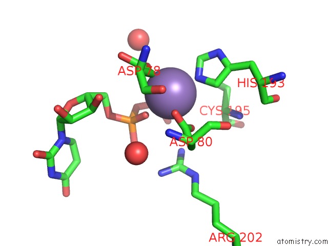
Mono view
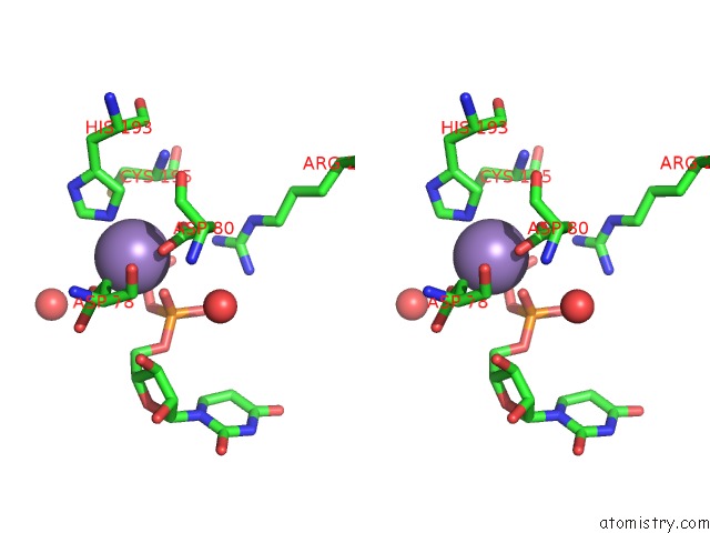
Stereo pair view
Mono view
Stereo pair view
A full contact list of Manganese with other atoms in the Mn binding
site number 1 of Crystal Structure of A Glycosyltransferase Involved in the Glycosylation of the Major Capsid of Pbcv-1 within 5.0Å range:
|
Reference:
Y.Zhang,
Y.Xiang,
J.L.Van Etten,
M.G.Rossmann.
Structure and Function of A Chlorella Virus-Encoded Glycosyltransferase. Structure V. 15 1031 2007.
ISSN: ISSN 0969-2126
PubMed: 17850743
DOI: 10.1016/J.STR.2007.07.006
Page generated: Sat Oct 5 14:50:34 2024
ISSN: ISSN 0969-2126
PubMed: 17850743
DOI: 10.1016/J.STR.2007.07.006
Last articles
Zn in 9J0NZn in 9J0O
Zn in 9J0P
Zn in 9FJX
Zn in 9EKB
Zn in 9C0F
Zn in 9CAH
Zn in 9CH0
Zn in 9CH3
Zn in 9CH1