Manganese »
PDB 2nzd-2p9a »
2ocl »
Manganese in PDB 2ocl: Crystal Structure of Valacyclovir Hydrolase S122A Mutant
Protein crystallography data
The structure of Crystal Structure of Valacyclovir Hydrolase S122A Mutant, PDB code: 2ocl
was solved by
L.Lai,
Z.Xu,
G.L.Amidon,
with X-Ray Crystallography technique. A brief refinement statistics is given in the table below:
| Resolution Low / High (Å) | 44.39 / 1.90 |
| Space group | P 62 |
| Cell size a, b, c (Å), α, β, γ (°) | 88.790, 88.790, 86.010, 90.00, 90.00, 120.00 |
| R / Rfree (%) | 18.4 / 20.8 |
Other elements in 2ocl:
The structure of Crystal Structure of Valacyclovir Hydrolase S122A Mutant also contains other interesting chemical elements:
| Magnesium | (Mg) | 1 atom |
Manganese Binding Sites:
The binding sites of Manganese atom in the Crystal Structure of Valacyclovir Hydrolase S122A Mutant
(pdb code 2ocl). This binding sites where shown within
5.0 Angstroms radius around Manganese atom.
In total only one binding site of Manganese was determined in the Crystal Structure of Valacyclovir Hydrolase S122A Mutant, PDB code: 2ocl:
In total only one binding site of Manganese was determined in the Crystal Structure of Valacyclovir Hydrolase S122A Mutant, PDB code: 2ocl:
Manganese binding site 1 out of 1 in 2ocl
Go back to
Manganese binding site 1 out
of 1 in the Crystal Structure of Valacyclovir Hydrolase S122A Mutant
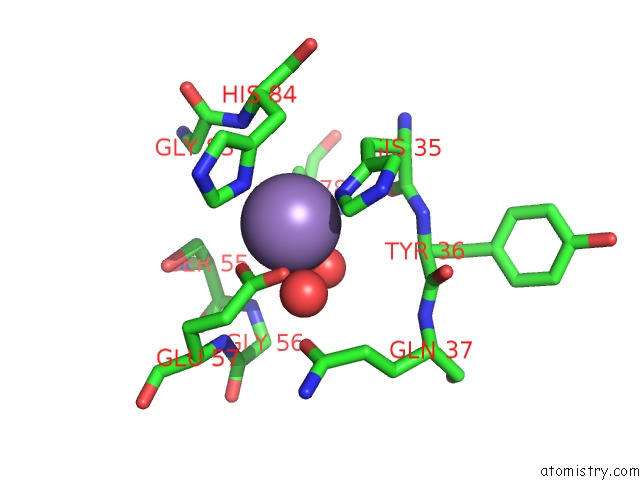
Mono view
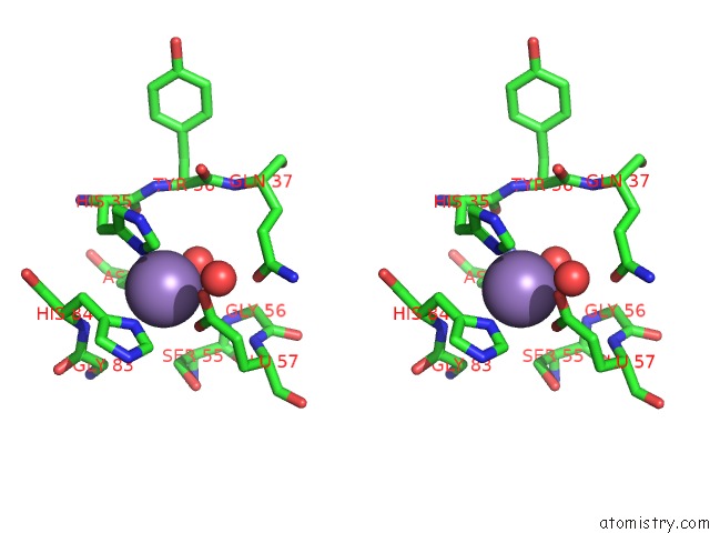
Stereo pair view
Mono view
Stereo pair view
A full contact list of Manganese with other atoms in the Mn binding
site number 1 of Crystal Structure of Valacyclovir Hydrolase S122A Mutant within 5.0Å range:
|
Reference:
L.Lai,
Z.Xu,
J.Zhou,
K.D.Lee,
G.L.Amidon.
Molecular Basis of Prodrug Activation By Human Valacyclovirase, An Alpha-Amino Acid Ester Hydrolase. J.Biol.Chem. V. 283 9318 2008.
ISSN: ISSN 0021-9258
PubMed: 18256025
DOI: 10.1074/JBC.M709530200
Page generated: Sat Oct 5 14:45:40 2024
ISSN: ISSN 0021-9258
PubMed: 18256025
DOI: 10.1074/JBC.M709530200
Last articles
Zn in 9J0NZn in 9J0O
Zn in 9J0P
Zn in 9FJX
Zn in 9EKB
Zn in 9C0F
Zn in 9CAH
Zn in 9CH0
Zn in 9CH3
Zn in 9CH1