Manganese »
PDB 2nzd-2p9a »
2o5q »
Manganese in PDB 2o5q: Manganese Horse Heart Myoglobin, Nitric Oxide Modified
Protein crystallography data
The structure of Manganese Horse Heart Myoglobin, Nitric Oxide Modified, PDB code: 2o5q
was solved by
G.B.Richter-Addo,
Z.N.Zahran,
L.Chooback,
D.M.Copeland,
A.H.West,
with X-Ray Crystallography technique. A brief refinement statistics is given in the table below:
| Resolution Low / High (Å) | 26.57 / 1.90 |
| Space group | P 1 21 1 |
| Cell size a, b, c (Å), α, β, γ (°) | 35.468, 28.637, 63.193, 90.00, 105.70, 90.00 |
| R / Rfree (%) | 17.7 / 23.5 |
Manganese Binding Sites:
The binding sites of Manganese atom in the Manganese Horse Heart Myoglobin, Nitric Oxide Modified
(pdb code 2o5q). This binding sites where shown within
5.0 Angstroms radius around Manganese atom.
In total only one binding site of Manganese was determined in the Manganese Horse Heart Myoglobin, Nitric Oxide Modified, PDB code: 2o5q:
In total only one binding site of Manganese was determined in the Manganese Horse Heart Myoglobin, Nitric Oxide Modified, PDB code: 2o5q:
Manganese binding site 1 out of 1 in 2o5q
Go back to
Manganese binding site 1 out
of 1 in the Manganese Horse Heart Myoglobin, Nitric Oxide Modified
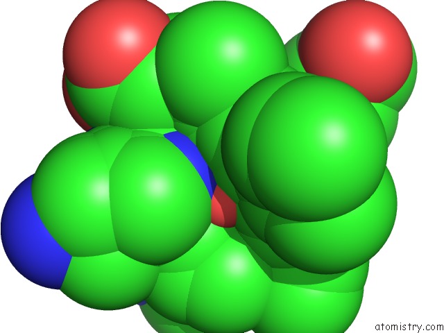
Mono view
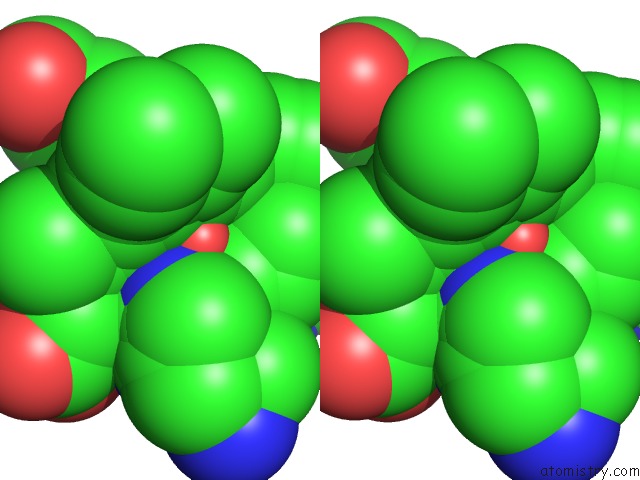
Stereo pair view
Mono view
Stereo pair view
A full contact list of Manganese with other atoms in the Mn binding
site number 1 of Manganese Horse Heart Myoglobin, Nitric Oxide Modified within 5.0Å range:
|
Reference:
Z.N.Zahran,
L.Chooback,
D.M.Copeland,
A.H.West,
G.B.Richter-Addo.
Crystal Structures of Manganese- and Cobalt-Substituted Myoglobin in Complex with No and Nitrite Reveal Unusual Ligand Conformations. J.Inorg.Biochem. V. 102 216 2008.
ISSN: ISSN 0162-0134
PubMed: 17905436
DOI: 10.1016/J.JINORGBIO.2007.08.002
Page generated: Sat Oct 5 14:44:50 2024
ISSN: ISSN 0162-0134
PubMed: 17905436
DOI: 10.1016/J.JINORGBIO.2007.08.002
Last articles
Zn in 9J0NZn in 9J0O
Zn in 9J0P
Zn in 9FJX
Zn in 9EKB
Zn in 9C0F
Zn in 9CAH
Zn in 9CH0
Zn in 9CH3
Zn in 9CH1