Manganese »
PDB 2jck-2nym »
2nw8 »
Manganese in PDB 2nw8: Crystal Structure of Tryptophan 2,3-Dioxygenase (Tdo) From Xanthomonas Campestris in Complex with Ferrous Heme and Tryptophan. Northeast Structural Genomics Target XCR13.
Protein crystallography data
The structure of Crystal Structure of Tryptophan 2,3-Dioxygenase (Tdo) From Xanthomonas Campestris in Complex with Ferrous Heme and Tryptophan. Northeast Structural Genomics Target XCR13., PDB code: 2nw8
was solved by
F.Forouhar,
J.L.R.Anderson,
C.G.Mowat,
C.Bruckmann,
S.J.Thackray,
J.Seetharaman,
C.K.Ho,
L.C.Ma,
K.Cunningham,
H.Janjua,
L.Zhao,
R.Xiao,
J.Liu,
M.C.Baran,
T.B.Acton,
B.Rost,
G.T.Montelione,
S.K.Champman,
L.Tong,
Northeast Structural Genomics Consortium (Nesg),
with X-Ray Crystallography technique. A brief refinement statistics is given in the table below:
| Resolution Low / High (Å) | 19.68 / 1.60 |
| Space group | P 31 2 1 |
| Cell size a, b, c (Å), α, β, γ (°) | 114.093, 114.094, 96.227, 90.00, 90.00, 120.00 |
| R / Rfree (%) | 17.1 / 18.9 |
Other elements in 2nw8:
The structure of Crystal Structure of Tryptophan 2,3-Dioxygenase (Tdo) From Xanthomonas Campestris in Complex with Ferrous Heme and Tryptophan. Northeast Structural Genomics Target XCR13. also contains other interesting chemical elements:
| Iron | (Fe) | 2 atoms |
Manganese Binding Sites:
The binding sites of Manganese atom in the Crystal Structure of Tryptophan 2,3-Dioxygenase (Tdo) From Xanthomonas Campestris in Complex with Ferrous Heme and Tryptophan. Northeast Structural Genomics Target XCR13.
(pdb code 2nw8). This binding sites where shown within
5.0 Angstroms radius around Manganese atom.
In total 2 binding sites of Manganese where determined in the Crystal Structure of Tryptophan 2,3-Dioxygenase (Tdo) From Xanthomonas Campestris in Complex with Ferrous Heme and Tryptophan. Northeast Structural Genomics Target XCR13., PDB code: 2nw8:
Jump to Manganese binding site number: 1; 2;
In total 2 binding sites of Manganese where determined in the Crystal Structure of Tryptophan 2,3-Dioxygenase (Tdo) From Xanthomonas Campestris in Complex with Ferrous Heme and Tryptophan. Northeast Structural Genomics Target XCR13., PDB code: 2nw8:
Jump to Manganese binding site number: 1; 2;
Manganese binding site 1 out of 2 in 2nw8
Go back to
Manganese binding site 1 out
of 2 in the Crystal Structure of Tryptophan 2,3-Dioxygenase (Tdo) From Xanthomonas Campestris in Complex with Ferrous Heme and Tryptophan. Northeast Structural Genomics Target XCR13.
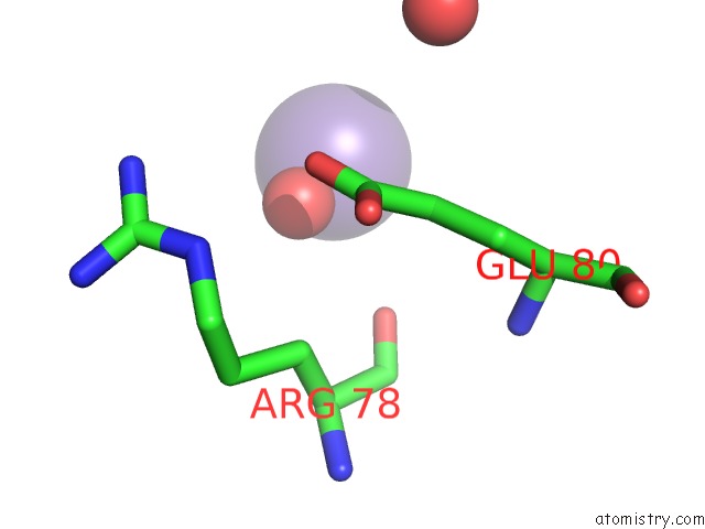
Mono view
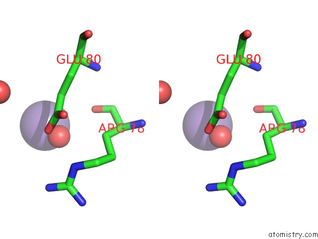
Stereo pair view
Mono view
Stereo pair view
A full contact list of Manganese with other atoms in the Mn binding
site number 1 of Crystal Structure of Tryptophan 2,3-Dioxygenase (Tdo) From Xanthomonas Campestris in Complex with Ferrous Heme and Tryptophan. Northeast Structural Genomics Target XCR13. within 5.0Å range:
|
Manganese binding site 2 out of 2 in 2nw8
Go back to
Manganese binding site 2 out
of 2 in the Crystal Structure of Tryptophan 2,3-Dioxygenase (Tdo) From Xanthomonas Campestris in Complex with Ferrous Heme and Tryptophan. Northeast Structural Genomics Target XCR13.
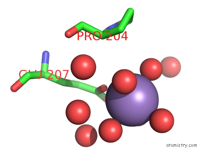
Mono view
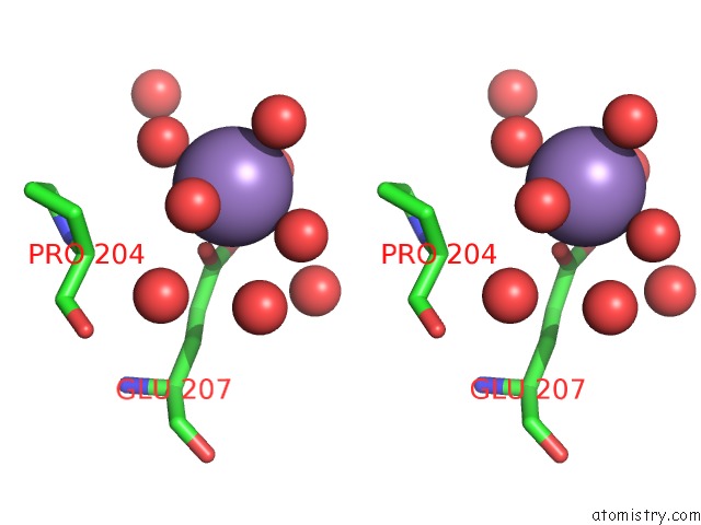
Stereo pair view
Mono view
Stereo pair view
A full contact list of Manganese with other atoms in the Mn binding
site number 2 of Crystal Structure of Tryptophan 2,3-Dioxygenase (Tdo) From Xanthomonas Campestris in Complex with Ferrous Heme and Tryptophan. Northeast Structural Genomics Target XCR13. within 5.0Å range:
|
Reference:
F.Forouhar,
J.L.Anderson,
C.G.Mowat,
S.M.Vorobiev,
A.Hussain,
M.Abashidze,
C.Bruckmann,
S.J.Thackray,
J.Seetharaman,
T.Tucker,
R.Xiao,
L.C.Ma,
L.Zhao,
T.B.Acton,
G.T.Montelione,
S.K.Chapman,
L.Tong.
Molecular Insights Into Substrate Recognition and Catalysis By Tryptophan 2,3-Dioxygenase. Proc.Natl.Acad.Sci.Usa V. 104 473 2007.
ISSN: ISSN 0027-8424
PubMed: 17197414
DOI: 10.1073/PNAS.0610007104
Page generated: Sat Oct 5 14:39:42 2024
ISSN: ISSN 0027-8424
PubMed: 17197414
DOI: 10.1073/PNAS.0610007104
Last articles
Zn in 9J0NZn in 9J0O
Zn in 9J0P
Zn in 9FJX
Zn in 9EKB
Zn in 9C0F
Zn in 9CAH
Zn in 9CH0
Zn in 9CH3
Zn in 9CH1