Manganese »
PDB 2jck-2nym »
2jla »
Manganese in PDB 2jla: Crystal Structure of E.Coli Mend, 2-Succinyl-5-Enolpyruvyl- 6-Hydroxy-3-Cyclohexadiene-1-Carboxylate Synthase - Semet Protein
Enzymatic activity of Crystal Structure of E.Coli Mend, 2-Succinyl-5-Enolpyruvyl- 6-Hydroxy-3-Cyclohexadiene-1-Carboxylate Synthase - Semet Protein
All present enzymatic activity of Crystal Structure of E.Coli Mend, 2-Succinyl-5-Enolpyruvyl- 6-Hydroxy-3-Cyclohexadiene-1-Carboxylate Synthase - Semet Protein:
2.2.1.9;
2.2.1.9;
Protein crystallography data
The structure of Crystal Structure of E.Coli Mend, 2-Succinyl-5-Enolpyruvyl- 6-Hydroxy-3-Cyclohexadiene-1-Carboxylate Synthase - Semet Protein, PDB code: 2jla
was solved by
A.Dawson,
P.K.Fyfe,
W.N.Hunter,
with X-Ray Crystallography technique. A brief refinement statistics is given in the table below:
| Resolution Low / High (Å) | 83.05 / 2.81 |
| Space group | P 65 |
| Cell size a, b, c (Å), α, β, γ (°) | 95.830, 95.830, 463.170, 90.00, 90.00, 120.00 |
| R / Rfree (%) | 16.9 / 23.3 |
Other elements in 2jla:
The structure of Crystal Structure of E.Coli Mend, 2-Succinyl-5-Enolpyruvyl- 6-Hydroxy-3-Cyclohexadiene-1-Carboxylate Synthase - Semet Protein also contains other interesting chemical elements:
| Chlorine | (Cl) | 13 atoms |
Manganese Binding Sites:
The binding sites of Manganese atom in the Crystal Structure of E.Coli Mend, 2-Succinyl-5-Enolpyruvyl- 6-Hydroxy-3-Cyclohexadiene-1-Carboxylate Synthase - Semet Protein
(pdb code 2jla). This binding sites where shown within
5.0 Angstroms radius around Manganese atom.
In total 5 binding sites of Manganese where determined in the Crystal Structure of E.Coli Mend, 2-Succinyl-5-Enolpyruvyl- 6-Hydroxy-3-Cyclohexadiene-1-Carboxylate Synthase - Semet Protein, PDB code: 2jla:
Jump to Manganese binding site number: 1; 2; 3; 4; 5;
In total 5 binding sites of Manganese where determined in the Crystal Structure of E.Coli Mend, 2-Succinyl-5-Enolpyruvyl- 6-Hydroxy-3-Cyclohexadiene-1-Carboxylate Synthase - Semet Protein, PDB code: 2jla:
Jump to Manganese binding site number: 1; 2; 3; 4; 5;
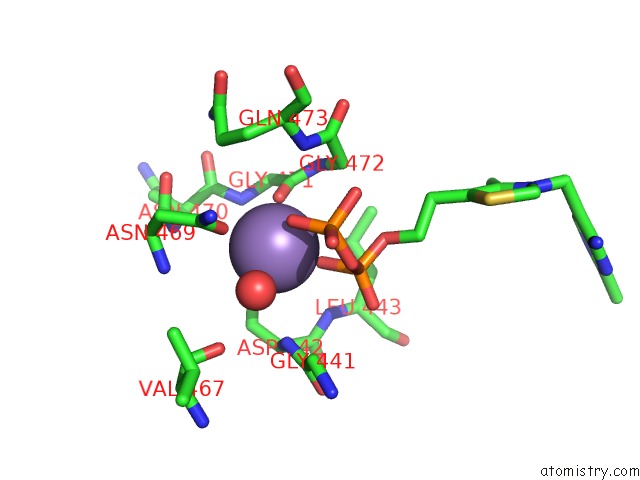
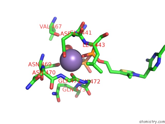
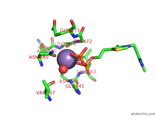
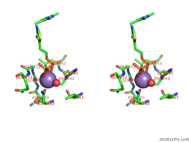
Manganese binding site 1 out of 5 in 2jla
Go back to
Manganese binding site 1 out
of 5 in the Crystal Structure of E.Coli Mend, 2-Succinyl-5-Enolpyruvyl- 6-Hydroxy-3-Cyclohexadiene-1-Carboxylate Synthase - Semet Protein
Mono view
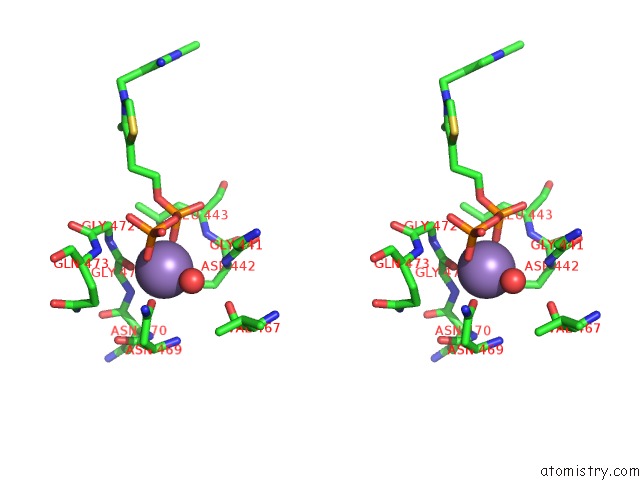
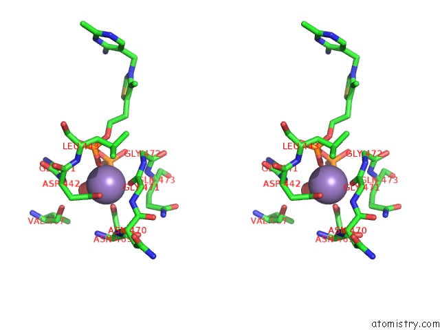
Stereo pair view
Mono view
Stereo pair view
A full contact list of Manganese with other atoms in the Mn binding
site number 1 of Crystal Structure of E.Coli Mend, 2-Succinyl-5-Enolpyruvyl- 6-Hydroxy-3-Cyclohexadiene-1-Carboxylate Synthase - Semet Protein within 5.0Å range:
|
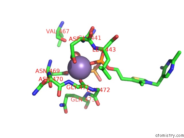
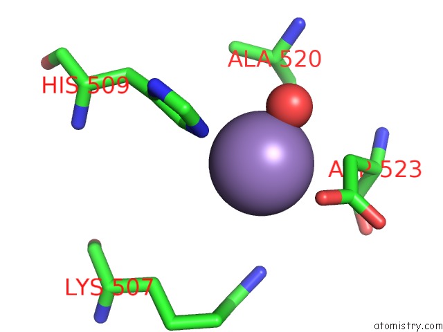
Manganese binding site 2 out of 5 in 2jla
Go back to
Manganese binding site 2 out
of 5 in the Crystal Structure of E.Coli Mend, 2-Succinyl-5-Enolpyruvyl- 6-Hydroxy-3-Cyclohexadiene-1-Carboxylate Synthase - Semet Protein
Mono view
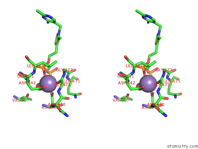
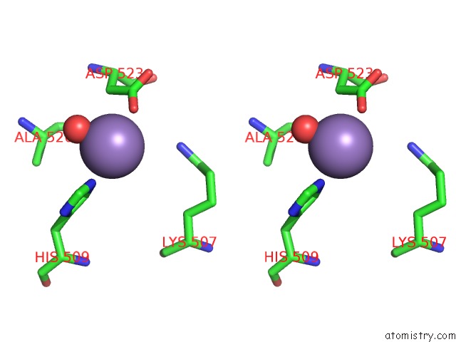
Stereo pair view
Mono view
Stereo pair view
A full contact list of Manganese with other atoms in the Mn binding
site number 2 of Crystal Structure of E.Coli Mend, 2-Succinyl-5-Enolpyruvyl- 6-Hydroxy-3-Cyclohexadiene-1-Carboxylate Synthase - Semet Protein within 5.0Å range:
|
Manganese binding site 3 out of 5 in 2jla
Go back to
Manganese binding site 3 out
of 5 in the Crystal Structure of E.Coli Mend, 2-Succinyl-5-Enolpyruvyl- 6-Hydroxy-3-Cyclohexadiene-1-Carboxylate Synthase - Semet Protein
Mono view
Stereo pair view
Mono view
Stereo pair view
A full contact list of Manganese with other atoms in the Mn binding
site number 3 of Crystal Structure of E.Coli Mend, 2-Succinyl-5-Enolpyruvyl- 6-Hydroxy-3-Cyclohexadiene-1-Carboxylate Synthase - Semet Protein within 5.0Å range:
|
Manganese binding site 4 out of 5 in 2jla
Go back to
Manganese binding site 4 out
of 5 in the Crystal Structure of E.Coli Mend, 2-Succinyl-5-Enolpyruvyl- 6-Hydroxy-3-Cyclohexadiene-1-Carboxylate Synthase - Semet Protein
Mono view
Stereo pair view
Mono view
Stereo pair view
A full contact list of Manganese with other atoms in the Mn binding
site number 4 of Crystal Structure of E.Coli Mend, 2-Succinyl-5-Enolpyruvyl- 6-Hydroxy-3-Cyclohexadiene-1-Carboxylate Synthase - Semet Protein within 5.0Å range:
|
Manganese binding site 5 out of 5 in 2jla
Go back to
Manganese binding site 5 out
of 5 in the Crystal Structure of E.Coli Mend, 2-Succinyl-5-Enolpyruvyl- 6-Hydroxy-3-Cyclohexadiene-1-Carboxylate Synthase - Semet Protein
Mono view
Stereo pair view
Mono view
Stereo pair view
A full contact list of Manganese with other atoms in the Mn binding
site number 5 of Crystal Structure of E.Coli Mend, 2-Succinyl-5-Enolpyruvyl- 6-Hydroxy-3-Cyclohexadiene-1-Carboxylate Synthase - Semet Protein within 5.0Å range:
|
Reference:
A.Dawson,
P.K.Fyfe,
W.N.Hunter.
Specificity and Reactivity in Menaquinone Biosynthesis: the Structure of Escherichia Coli Mend (2-Succinyl-5-Enolpyruvyl-6-Hydroxy-3- Cyclohexadiene-1-Carboxylate Synthase). J.Mol.Biol. V. 384 1353 2008.
ISSN: ISSN 0022-2836
PubMed: 18983854
DOI: 10.1016/J.JMB.2008.10.048
Page generated: Sat Oct 5 14:35:22 2024
ISSN: ISSN 0022-2836
PubMed: 18983854
DOI: 10.1016/J.JMB.2008.10.048
Last articles
F in 7L7OF in 7L5E
F in 7L72
F in 7L5P
F in 7L69
F in 7L5O
F in 7L0K
F in 7L4W
F in 7L4U
F in 7L4N