Manganese »
PDB 2hvh-2jcj »
2iae »
Manganese in PDB 2iae: Crystal Structure of A Protein Phosphatase 2A (PP2A) Holoenzyme.
Enzymatic activity of Crystal Structure of A Protein Phosphatase 2A (PP2A) Holoenzyme.
All present enzymatic activity of Crystal Structure of A Protein Phosphatase 2A (PP2A) Holoenzyme.:
3.1.3.16;
3.1.3.16;
Protein crystallography data
The structure of Crystal Structure of A Protein Phosphatase 2A (PP2A) Holoenzyme., PDB code: 2iae
was solved by
U.S.Cho,
W.Xu,
with X-Ray Crystallography technique. A brief refinement statistics is given in the table below:
| Resolution Low / High (Å) | 20.00 / 3.50 |
| Space group | P 21 3 |
| Cell size a, b, c (Å), α, β, γ (°) | 265.301, 265.301, 265.301, 90.00, 90.00, 90.00 |
| R / Rfree (%) | 25.7 / 31.6 |
Manganese Binding Sites:
The binding sites of Manganese atom in the Crystal Structure of A Protein Phosphatase 2A (PP2A) Holoenzyme.
(pdb code 2iae). This binding sites where shown within
5.0 Angstroms radius around Manganese atom.
In total 4 binding sites of Manganese where determined in the Crystal Structure of A Protein Phosphatase 2A (PP2A) Holoenzyme., PDB code: 2iae:
Jump to Manganese binding site number: 1; 2; 3; 4;
In total 4 binding sites of Manganese where determined in the Crystal Structure of A Protein Phosphatase 2A (PP2A) Holoenzyme., PDB code: 2iae:
Jump to Manganese binding site number: 1; 2; 3; 4;
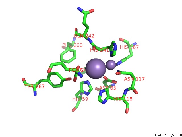
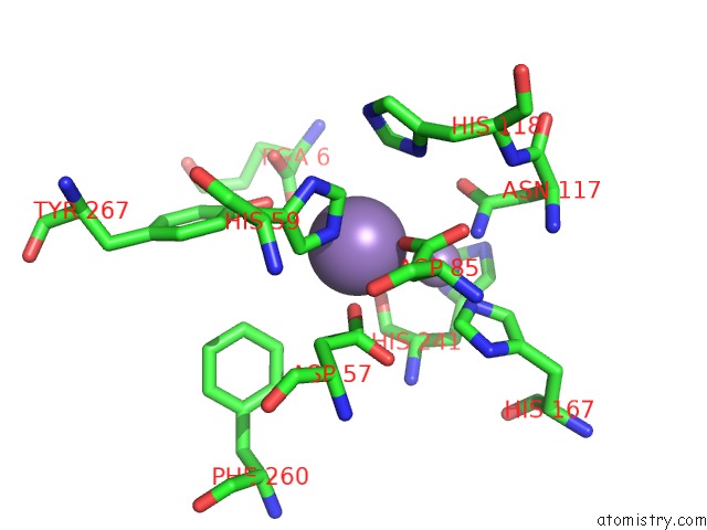
Manganese binding site 1 out of 4 in 2iae
Go back to
Manganese binding site 1 out
of 4 in the Crystal Structure of A Protein Phosphatase 2A (PP2A) Holoenzyme.
Mono view
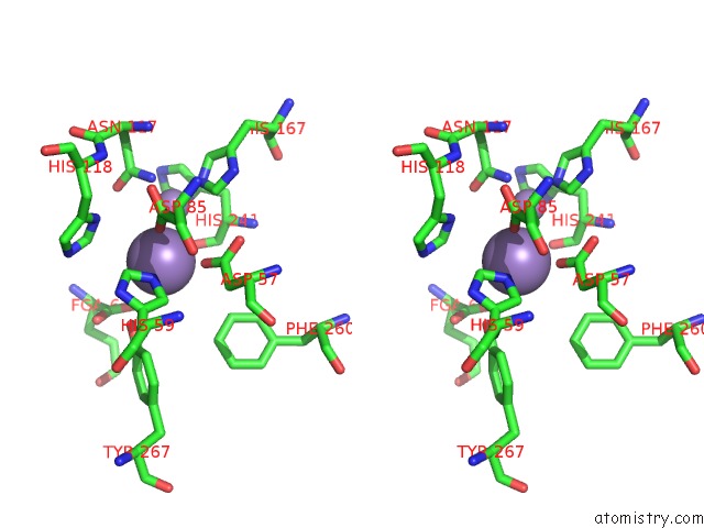
Stereo pair view
Mono view
Stereo pair view
A full contact list of Manganese with other atoms in the Mn binding
site number 1 of Crystal Structure of A Protein Phosphatase 2A (PP2A) Holoenzyme. within 5.0Å range:
|
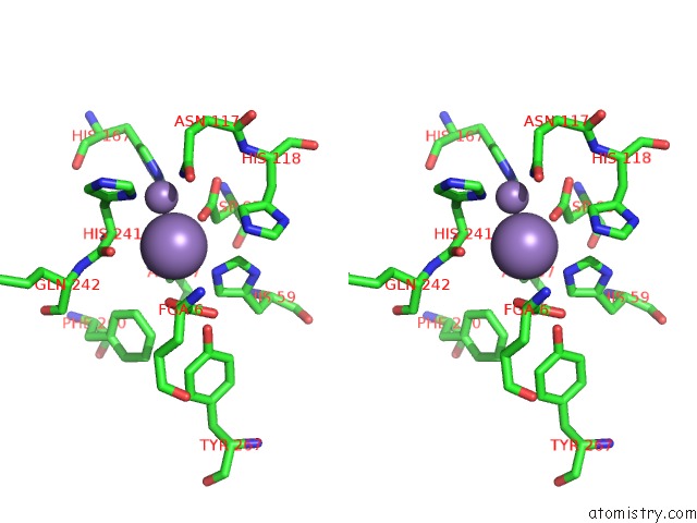
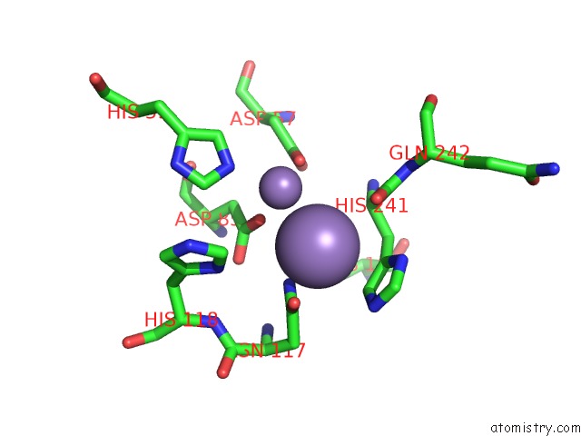
Manganese binding site 2 out of 4 in 2iae
Go back to
Manganese binding site 2 out
of 4 in the Crystal Structure of A Protein Phosphatase 2A (PP2A) Holoenzyme.
Mono view
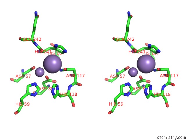
Stereo pair view
Mono view
Stereo pair view
A full contact list of Manganese with other atoms in the Mn binding
site number 2 of Crystal Structure of A Protein Phosphatase 2A (PP2A) Holoenzyme. within 5.0Å range:
|
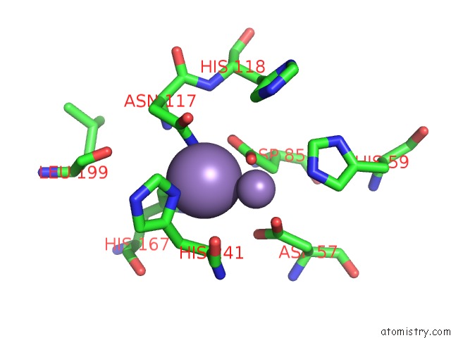
Manganese binding site 3 out of 4 in 2iae
Go back to
Manganese binding site 3 out
of 4 in the Crystal Structure of A Protein Phosphatase 2A (PP2A) Holoenzyme.
Mono view
Stereo pair view
Mono view
Stereo pair view
A full contact list of Manganese with other atoms in the Mn binding
site number 3 of Crystal Structure of A Protein Phosphatase 2A (PP2A) Holoenzyme. within 5.0Å range:
|
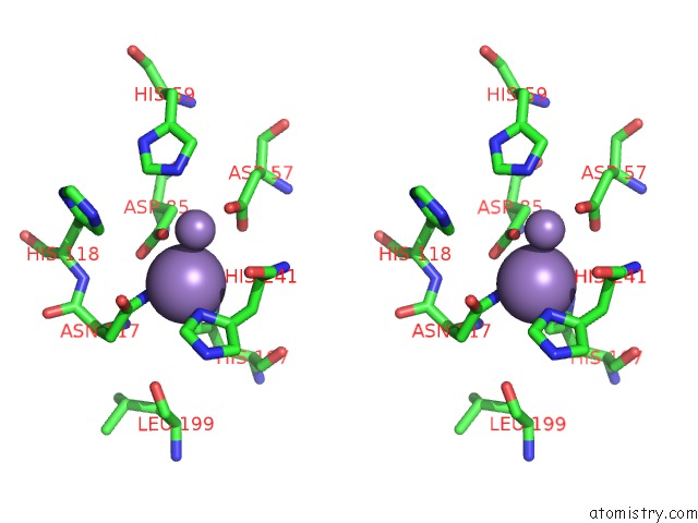
Manganese binding site 4 out of 4 in 2iae
Go back to
Manganese binding site 4 out
of 4 in the Crystal Structure of A Protein Phosphatase 2A (PP2A) Holoenzyme.
Mono view
Stereo pair view
Mono view
Stereo pair view
A full contact list of Manganese with other atoms in the Mn binding
site number 4 of Crystal Structure of A Protein Phosphatase 2A (PP2A) Holoenzyme. within 5.0Å range:
|
Reference:
U.S.Cho,
W.Xu.
Crystal Structure of A Protein Phosphatase 2A Heterotrimeric Holoenzyme. Nature V. 445 53 2007.
ISSN: ISSN 0028-0836
PubMed: 17086192
DOI: 10.1038/NATURE05351
Page generated: Sat Oct 5 14:26:20 2024
ISSN: ISSN 0028-0836
PubMed: 17086192
DOI: 10.1038/NATURE05351
Last articles
Zn in 9MJ5Zn in 9HNW
Zn in 9G0L
Zn in 9FNE
Zn in 9DZN
Zn in 9E0I
Zn in 9D32
Zn in 9DAK
Zn in 8ZXC
Zn in 8ZUF