Manganese »
PDB 2feu-2hr6 »
2hr6 »
Manganese in PDB 2hr6: Crystal Structure of Dutpase in Complex with Substrate Analogue Dudp and Manganese
Enzymatic activity of Crystal Structure of Dutpase in Complex with Substrate Analogue Dudp and Manganese
All present enzymatic activity of Crystal Structure of Dutpase in Complex with Substrate Analogue Dudp and Manganese:
3.6.1.23;
3.6.1.23;
Protein crystallography data
The structure of Crystal Structure of Dutpase in Complex with Substrate Analogue Dudp and Manganese, PDB code: 2hr6
was solved by
O.Barabas,
J.Kovari,
R.Tapai,
B.G.Vertessy,
with X-Ray Crystallography technique. A brief refinement statistics is given in the table below:
| Resolution Low / High (Å) | 24.90 / 1.84 |
| Space group | P 63 2 2 |
| Cell size a, b, c (Å), α, β, γ (°) | 75.049, 75.049, 99.936, 90.00, 90.00, 120.00 |
| R / Rfree (%) | 15.1 / 17.3 |
Manganese Binding Sites:
The binding sites of Manganese atom in the Crystal Structure of Dutpase in Complex with Substrate Analogue Dudp and Manganese
(pdb code 2hr6). This binding sites where shown within
5.0 Angstroms radius around Manganese atom.
In total only one binding site of Manganese was determined in the Crystal Structure of Dutpase in Complex with Substrate Analogue Dudp and Manganese, PDB code: 2hr6:
In total only one binding site of Manganese was determined in the Crystal Structure of Dutpase in Complex with Substrate Analogue Dudp and Manganese, PDB code: 2hr6:
Manganese binding site 1 out of 1 in 2hr6
Go back to
Manganese binding site 1 out
of 1 in the Crystal Structure of Dutpase in Complex with Substrate Analogue Dudp and Manganese
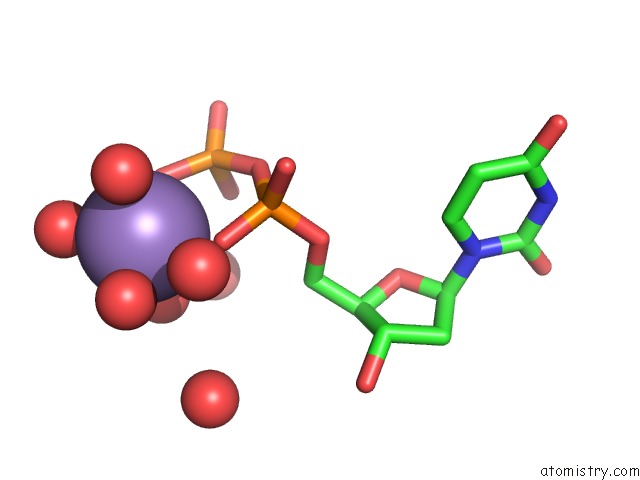
Mono view
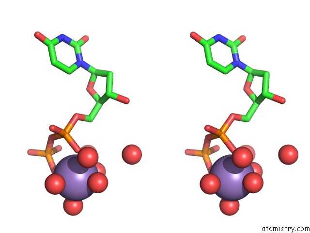
Stereo pair view
Mono view
Stereo pair view
A full contact list of Manganese with other atoms in the Mn binding
site number 1 of Crystal Structure of Dutpase in Complex with Substrate Analogue Dudp and Manganese within 5.0Å range:
|
Reference:
J.Kovari,
O.Barabas,
B.Varga,
A.Bekesi,
F.Tolgyesi,
J.Fidy,
J.Nagy,
B.G.Vertessy.
Methylene Substitution at the Alpha-Beta Bridging Position Within the Phosphate Chain of Dudp Profoundly Perturbs Ligand Accommodation Into the Dutpase Active Site. Proteins V. 71 308 2008.
ISSN: ISSN 0887-3585
PubMed: 17932923
DOI: 10.1002/PROT.21757
Page generated: Sat Oct 5 14:18:33 2024
ISSN: ISSN 0887-3585
PubMed: 17932923
DOI: 10.1002/PROT.21757
Last articles
Zn in 9MJ5Zn in 9HNW
Zn in 9G0L
Zn in 9FNE
Zn in 9DZN
Zn in 9E0I
Zn in 9D32
Zn in 9DAK
Zn in 8ZXC
Zn in 8ZUF