Manganese »
PDB 2feu-2hr6 »
2fyd »
Manganese in PDB 2fyd: Catalytic Domain of Bovine Beta 1, 4-Galactosyltransferase in Complex with Alpha-Lactalbumin, Glucose, Mn, and Udp-N- Acetylgalactosamine
Protein crystallography data
The structure of Catalytic Domain of Bovine Beta 1, 4-Galactosyltransferase in Complex with Alpha-Lactalbumin, Glucose, Mn, and Udp-N- Acetylgalactosamine, PDB code: 2fyd
was solved by
B.Ramakrishnan,
V.Ramasamy,
P.K.Qasba,
with X-Ray Crystallography technique. A brief refinement statistics is given in the table below:
| Resolution Low / High (Å) | 32.08 / 2.00 |
| Space group | P 1 21 1 |
| Cell size a, b, c (Å), α, β, γ (°) | 58.068, 95.376, 100.389, 90.00, 101.33, 90.00 |
| R / Rfree (%) | 20.1 / 24.2 |
Other elements in 2fyd:
The structure of Catalytic Domain of Bovine Beta 1, 4-Galactosyltransferase in Complex with Alpha-Lactalbumin, Glucose, Mn, and Udp-N- Acetylgalactosamine also contains other interesting chemical elements:
| Calcium | (Ca) | 2 atoms |
Manganese Binding Sites:
The binding sites of Manganese atom in the Catalytic Domain of Bovine Beta 1, 4-Galactosyltransferase in Complex with Alpha-Lactalbumin, Glucose, Mn, and Udp-N- Acetylgalactosamine
(pdb code 2fyd). This binding sites where shown within
5.0 Angstroms radius around Manganese atom.
In total 2 binding sites of Manganese where determined in the Catalytic Domain of Bovine Beta 1, 4-Galactosyltransferase in Complex with Alpha-Lactalbumin, Glucose, Mn, and Udp-N- Acetylgalactosamine, PDB code: 2fyd:
Jump to Manganese binding site number: 1; 2;
In total 2 binding sites of Manganese where determined in the Catalytic Domain of Bovine Beta 1, 4-Galactosyltransferase in Complex with Alpha-Lactalbumin, Glucose, Mn, and Udp-N- Acetylgalactosamine, PDB code: 2fyd:
Jump to Manganese binding site number: 1; 2;
Manganese binding site 1 out of 2 in 2fyd
Go back to
Manganese binding site 1 out
of 2 in the Catalytic Domain of Bovine Beta 1, 4-Galactosyltransferase in Complex with Alpha-Lactalbumin, Glucose, Mn, and Udp-N- Acetylgalactosamine
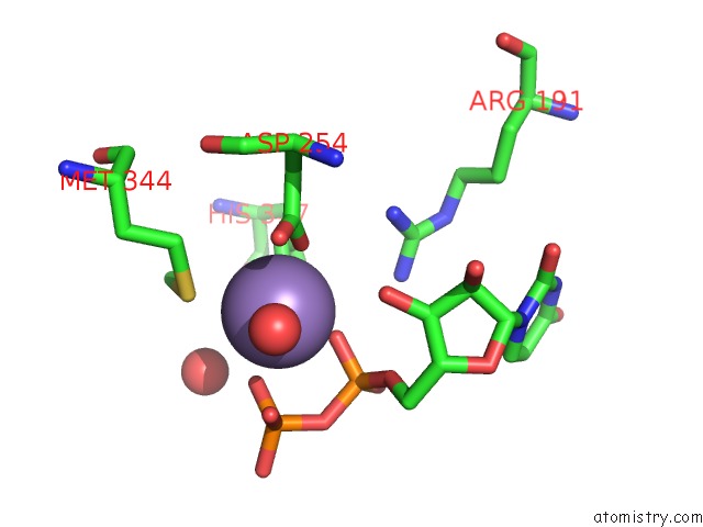
Mono view
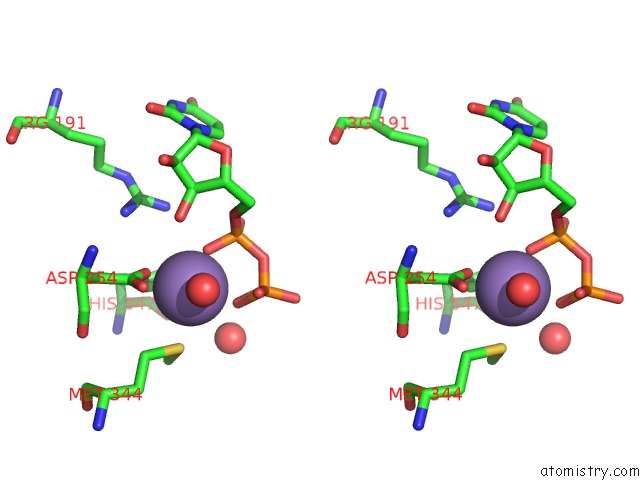
Stereo pair view
Mono view
Stereo pair view
A full contact list of Manganese with other atoms in the Mn binding
site number 1 of Catalytic Domain of Bovine Beta 1, 4-Galactosyltransferase in Complex with Alpha-Lactalbumin, Glucose, Mn, and Udp-N- Acetylgalactosamine within 5.0Å range:
|
Manganese binding site 2 out of 2 in 2fyd
Go back to
Manganese binding site 2 out
of 2 in the Catalytic Domain of Bovine Beta 1, 4-Galactosyltransferase in Complex with Alpha-Lactalbumin, Glucose, Mn, and Udp-N- Acetylgalactosamine
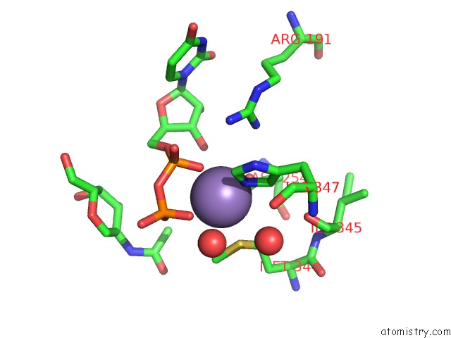
Mono view
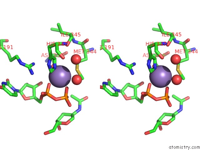
Stereo pair view
Mono view
Stereo pair view
A full contact list of Manganese with other atoms in the Mn binding
site number 2 of Catalytic Domain of Bovine Beta 1, 4-Galactosyltransferase in Complex with Alpha-Lactalbumin, Glucose, Mn, and Udp-N- Acetylgalactosamine within 5.0Å range:
|
Reference:
B.Ramakrishnan,
V.Ramasamy,
P.K.Qasba.
Structural Snapshots of Beta-1,4-Galactosyltransferase-I Along the Kinetic Pathway. J.Mol.Biol. V. 357 1619 2006.
ISSN: ISSN 0022-2836
PubMed: 16497331
DOI: 10.1016/J.JMB.2006.01.088
Page generated: Sat Oct 5 14:08:50 2024
ISSN: ISSN 0022-2836
PubMed: 16497331
DOI: 10.1016/J.JMB.2006.01.088
Last articles
Ca in 5SBDCa in 5SBE
Ca in 5SBC
Ca in 5SBB
Ca in 5SBA
Ca in 5SB7
Ca in 5SB9
Ca in 5SB8
Ca in 5SB6
Ca in 5SB5