Manganese »
PDB 2dvd-2fer »
2f1d »
Manganese in PDB 2f1d: X-Ray Structure of Imidazoleglycerol-Phosphate Dehydratase
Enzymatic activity of X-Ray Structure of Imidazoleglycerol-Phosphate Dehydratase
All present enzymatic activity of X-Ray Structure of Imidazoleglycerol-Phosphate Dehydratase:
4.2.1.19;
4.2.1.19;
Protein crystallography data
The structure of X-Ray Structure of Imidazoleglycerol-Phosphate Dehydratase, PDB code: 2f1d
was solved by
D.W.Rice,
S.E.Glynn,
P.J.Baker,
S.E.Sedelnikova,
C.L.Davies,
T.C.Eadsforth,
with X-Ray Crystallography technique. A brief refinement statistics is given in the table below:
| Resolution Low / High (Å) | 20.00 / 3.00 |
| Space group | H 3 1 |
| Cell size a, b, c (Å), α, β, γ (°) | 157.949, 157.949, 479.965, 90.00, 90.00, 120.00 |
| R / Rfree (%) | 24 / 28.6 |
Manganese Binding Sites:
Pages:
>>> Page 1 <<< Page 2, Binding sites: 11 - 20; Page 3, Binding sites: 21 - 30; Page 4, Binding sites: 31 - 32;Binding sites:
The binding sites of Manganese atom in the X-Ray Structure of Imidazoleglycerol-Phosphate Dehydratase (pdb code 2f1d). This binding sites where shown within 5.0 Angstroms radius around Manganese atom.In total 32 binding sites of Manganese where determined in the X-Ray Structure of Imidazoleglycerol-Phosphate Dehydratase, PDB code: 2f1d:
Jump to Manganese binding site number: 1; 2; 3; 4; 5; 6; 7; 8; 9; 10;
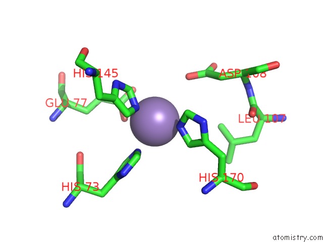
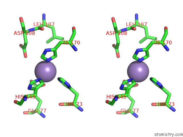
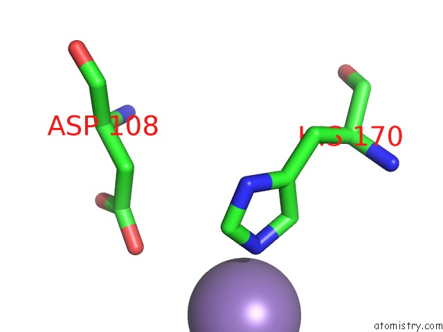
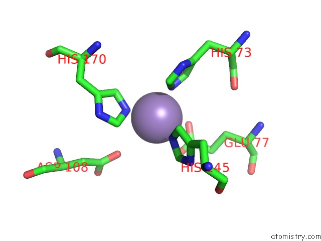
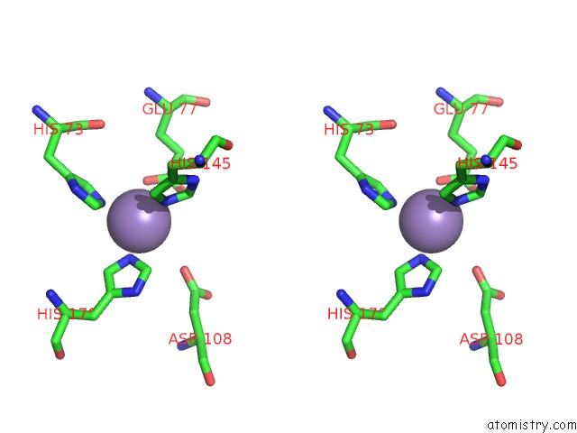
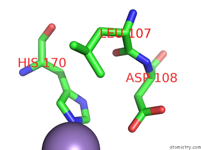
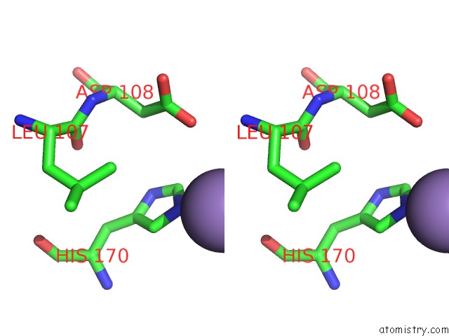
Manganese binding site 1 out of 32 in 2f1d
Go back to
Manganese binding site 1 out
of 32 in the X-Ray Structure of Imidazoleglycerol-Phosphate Dehydratase
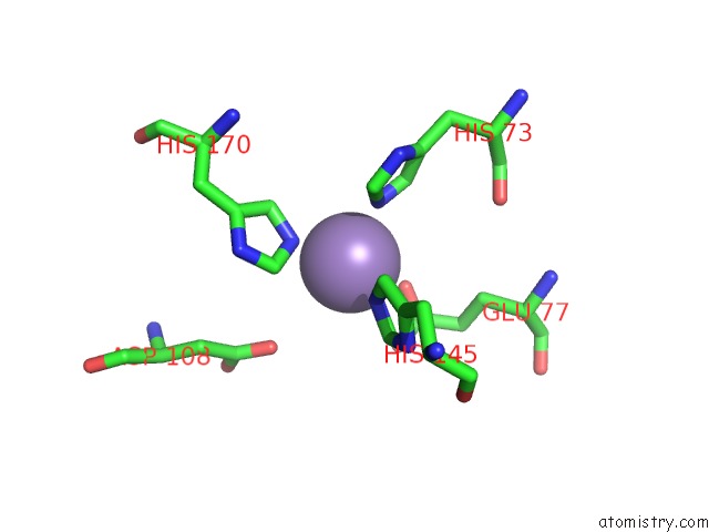
Mono view
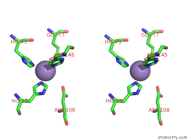
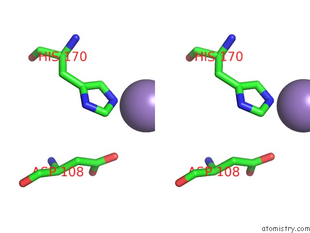
Stereo pair view
Mono view
Stereo pair view
A full contact list of Manganese with other atoms in the Mn binding
site number 1 of X-Ray Structure of Imidazoleglycerol-Phosphate Dehydratase within 5.0Å range:
|
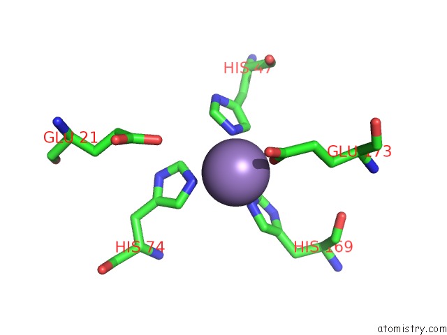
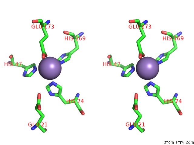
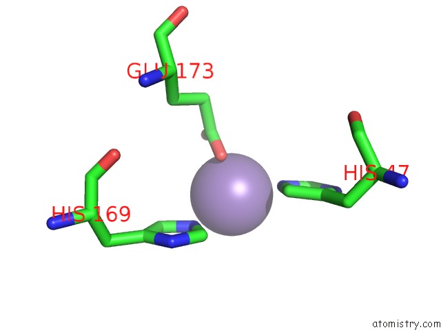
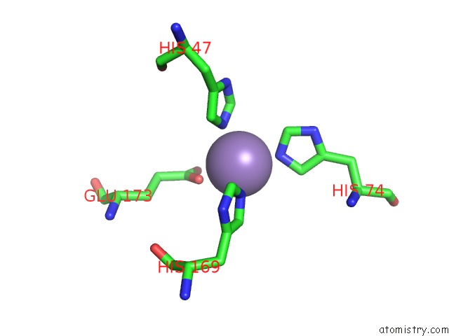
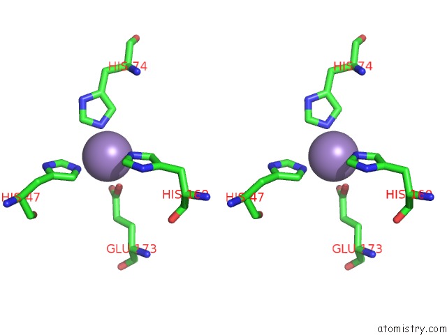
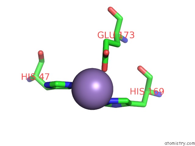
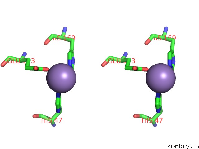
Manganese binding site 2 out of 32 in 2f1d
Go back to
Manganese binding site 2 out
of 32 in the X-Ray Structure of Imidazoleglycerol-Phosphate Dehydratase
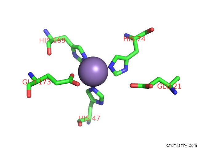
Mono view
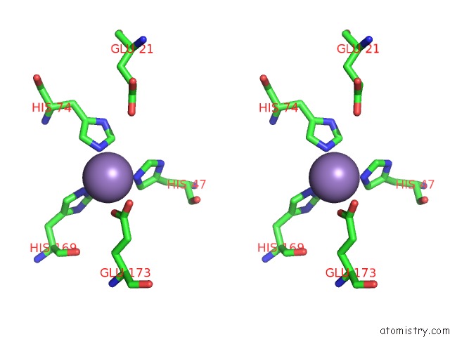
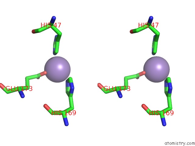
Stereo pair view
Mono view
Stereo pair view
A full contact list of Manganese with other atoms in the Mn binding
site number 2 of X-Ray Structure of Imidazoleglycerol-Phosphate Dehydratase within 5.0Å range:
|
Manganese binding site 3 out of 32 in 2f1d
Go back to
Manganese binding site 3 out
of 32 in the X-Ray Structure of Imidazoleglycerol-Phosphate Dehydratase
Mono view
Stereo pair view
Mono view
Stereo pair view
A full contact list of Manganese with other atoms in the Mn binding
site number 3 of X-Ray Structure of Imidazoleglycerol-Phosphate Dehydratase within 5.0Å range:
|
Manganese binding site 4 out of 32 in 2f1d
Go back to
Manganese binding site 4 out
of 32 in the X-Ray Structure of Imidazoleglycerol-Phosphate Dehydratase
Mono view
Stereo pair view
Mono view
Stereo pair view
A full contact list of Manganese with other atoms in the Mn binding
site number 4 of X-Ray Structure of Imidazoleglycerol-Phosphate Dehydratase within 5.0Å range:
|
Manganese binding site 5 out of 32 in 2f1d
Go back to
Manganese binding site 5 out
of 32 in the X-Ray Structure of Imidazoleglycerol-Phosphate Dehydratase
Mono view
Stereo pair view
Mono view
Stereo pair view
A full contact list of Manganese with other atoms in the Mn binding
site number 5 of X-Ray Structure of Imidazoleglycerol-Phosphate Dehydratase within 5.0Å range:
|
Manganese binding site 6 out of 32 in 2f1d
Go back to
Manganese binding site 6 out
of 32 in the X-Ray Structure of Imidazoleglycerol-Phosphate Dehydratase
Mono view
Stereo pair view
Mono view
Stereo pair view
A full contact list of Manganese with other atoms in the Mn binding
site number 6 of X-Ray Structure of Imidazoleglycerol-Phosphate Dehydratase within 5.0Å range:
|
Manganese binding site 7 out of 32 in 2f1d
Go back to
Manganese binding site 7 out
of 32 in the X-Ray Structure of Imidazoleglycerol-Phosphate Dehydratase
Mono view
Stereo pair view
Mono view
Stereo pair view
A full contact list of Manganese with other atoms in the Mn binding
site number 7 of X-Ray Structure of Imidazoleglycerol-Phosphate Dehydratase within 5.0Å range:
|
Manganese binding site 8 out of 32 in 2f1d
Go back to
Manganese binding site 8 out
of 32 in the X-Ray Structure of Imidazoleglycerol-Phosphate Dehydratase
Mono view
Stereo pair view
Mono view
Stereo pair view
A full contact list of Manganese with other atoms in the Mn binding
site number 8 of X-Ray Structure of Imidazoleglycerol-Phosphate Dehydratase within 5.0Å range:
|
Manganese binding site 9 out of 32 in 2f1d
Go back to
Manganese binding site 9 out
of 32 in the X-Ray Structure of Imidazoleglycerol-Phosphate Dehydratase
Mono view
Stereo pair view
Mono view
Stereo pair view
A full contact list of Manganese with other atoms in the Mn binding
site number 9 of X-Ray Structure of Imidazoleglycerol-Phosphate Dehydratase within 5.0Å range:
|
Manganese binding site 10 out of 32 in 2f1d
Go back to
Manganese binding site 10 out
of 32 in the X-Ray Structure of Imidazoleglycerol-Phosphate Dehydratase
Mono view
Stereo pair view
Mono view
Stereo pair view
A full contact list of Manganese with other atoms in the Mn binding
site number 10 of X-Ray Structure of Imidazoleglycerol-Phosphate Dehydratase within 5.0Å range:
|
Reference:
S.E.Glynn,
P.J.Baker,
S.E.Sedelnikova,
C.L.Davies,
T.C.Eadsforth,
C.W.Levy,
H.F.Rodgers,
G.M.Blackburn,
T.R.Hawkes,
R.Viner,
D.W.Rice.
Structure and Mechanism of Imidazoleglycerol-Phosphate Dehydratase. Structure V. 13 1809 2005.
ISSN: ISSN 0969-2126
PubMed: 16338409
DOI: 10.1016/J.STR.2005.08.012
Page generated: Sat Oct 5 14:01:10 2024
ISSN: ISSN 0969-2126
PubMed: 16338409
DOI: 10.1016/J.STR.2005.08.012
Last articles
Zn in 9MJ5Zn in 9HNW
Zn in 9G0L
Zn in 9FNE
Zn in 9DZN
Zn in 9E0I
Zn in 9D32
Zn in 9DAK
Zn in 8ZXC
Zn in 8ZUF