Manganese »
PDB 2dvd-2fer »
2et7 »
Manganese in PDB 2et7: Structural and Spectroscopic Insights Into the Mechanism of Oxalate Oxidase
Enzymatic activity of Structural and Spectroscopic Insights Into the Mechanism of Oxalate Oxidase
All present enzymatic activity of Structural and Spectroscopic Insights Into the Mechanism of Oxalate Oxidase:
1.2.3.4;
1.2.3.4;
Protein crystallography data
The structure of Structural and Spectroscopic Insights Into the Mechanism of Oxalate Oxidase, PDB code: 2et7
was solved by
O.Opaleye,
R.-S.Rose,
M.M.Whittaker,
E.-J.Woo,
J.W.Whittaker,
R.W.Pickersgill,
with X-Ray Crystallography technique. A brief refinement statistics is given in the table below:
| Resolution Low / High (Å) | 30.00 / 1.70 |
| Space group | H 3 2 |
| Cell size a, b, c (Å), α, β, γ (°) | 94.823, 94.823, 106.238, 90.00, 90.00, 120.00 |
| R / Rfree (%) | 16.3 / 21.1 |
Manganese Binding Sites:
The binding sites of Manganese atom in the Structural and Spectroscopic Insights Into the Mechanism of Oxalate Oxidase
(pdb code 2et7). This binding sites where shown within
5.0 Angstroms radius around Manganese atom.
In total only one binding site of Manganese was determined in the Structural and Spectroscopic Insights Into the Mechanism of Oxalate Oxidase, PDB code: 2et7:
In total only one binding site of Manganese was determined in the Structural and Spectroscopic Insights Into the Mechanism of Oxalate Oxidase, PDB code: 2et7:
Manganese binding site 1 out of 1 in 2et7
Go back to
Manganese binding site 1 out
of 1 in the Structural and Spectroscopic Insights Into the Mechanism of Oxalate Oxidase
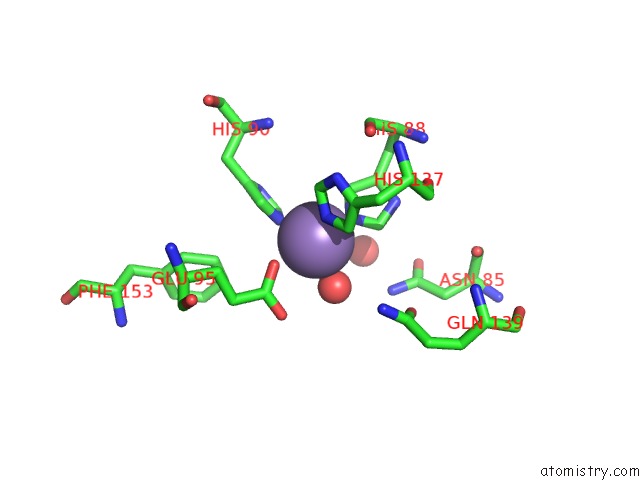
Mono view
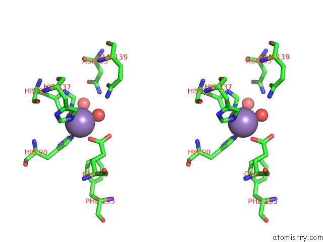
Stereo pair view
Mono view
Stereo pair view
A full contact list of Manganese with other atoms in the Mn binding
site number 1 of Structural and Spectroscopic Insights Into the Mechanism of Oxalate Oxidase within 5.0Å range:
|
Reference:
O.Opaleye,
R.-S.Rose,
M.M.Whittaker,
E.-J.Woo,
J.W.Whittaker,
R.W.Pickersgill.
Structural and Spectroscopic Studies Shed Light on the Mechanism of Oxalate Oxidase J.Biol.Chem. V. 281 6428 2006.
ISSN: ISSN 0021-9258
PubMed: 16291738
DOI: 10.1074/JBC.M510256200
Page generated: Sat Oct 5 13:59:50 2024
ISSN: ISSN 0021-9258
PubMed: 16291738
DOI: 10.1074/JBC.M510256200
Last articles
Ca in 3A7YCa in 3A7X
Ca in 3A7W
Ca in 3A7V
Ca in 3A7T
Ca in 3A7Q
Ca in 3A6O
Ca in 3A5N
Ca in 3A5O
Ca in 3A5M