Manganese »
PDB 2dvd-2fer »
2e7t »
Manganese in PDB 2e7t: Crystal Structure of Basic Winged Bean Lectin in Complex with A Blood Group Trisaccharide
Protein crystallography data
The structure of Crystal Structure of Basic Winged Bean Lectin in Complex with A Blood Group Trisaccharide, PDB code: 2e7t
was solved by
K.A.Kulkarni,
S.Katiyar,
A.Surolia,
M.Vijayan,
K.Suguna,
with X-Ray Crystallography technique. A brief refinement statistics is given in the table below:
| Resolution Low / High (Å) | 29.00 / 2.65 |
| Space group | P 21 21 2 |
| Cell size a, b, c (Å), α, β, γ (°) | 157.729, 91.130, 73.718, 90.00, 90.00, 90.00 |
| R / Rfree (%) | 19.2 / 24.4 |
Other elements in 2e7t:
The structure of Crystal Structure of Basic Winged Bean Lectin in Complex with A Blood Group Trisaccharide also contains other interesting chemical elements:
| Calcium | (Ca) | 4 atoms |
Manganese Binding Sites:
The binding sites of Manganese atom in the Crystal Structure of Basic Winged Bean Lectin in Complex with A Blood Group Trisaccharide
(pdb code 2e7t). This binding sites where shown within
5.0 Angstroms radius around Manganese atom.
In total 4 binding sites of Manganese where determined in the Crystal Structure of Basic Winged Bean Lectin in Complex with A Blood Group Trisaccharide, PDB code: 2e7t:
Jump to Manganese binding site number: 1; 2; 3; 4;
In total 4 binding sites of Manganese where determined in the Crystal Structure of Basic Winged Bean Lectin in Complex with A Blood Group Trisaccharide, PDB code: 2e7t:
Jump to Manganese binding site number: 1; 2; 3; 4;
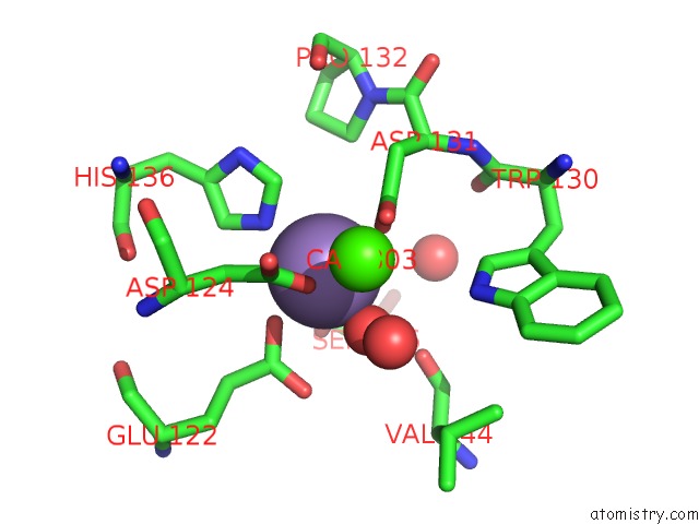
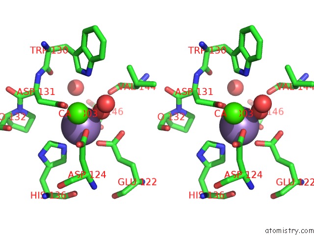
Manganese binding site 1 out of 4 in 2e7t
Go back to
Manganese binding site 1 out
of 4 in the Crystal Structure of Basic Winged Bean Lectin in Complex with A Blood Group Trisaccharide
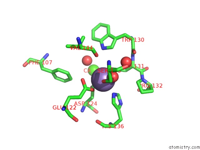
Mono view
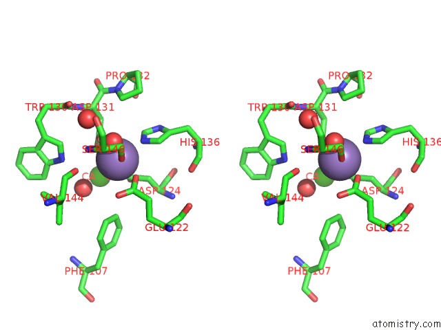
Stereo pair view
Mono view
Stereo pair view
A full contact list of Manganese with other atoms in the Mn binding
site number 1 of Crystal Structure of Basic Winged Bean Lectin in Complex with A Blood Group Trisaccharide within 5.0Å range:
|
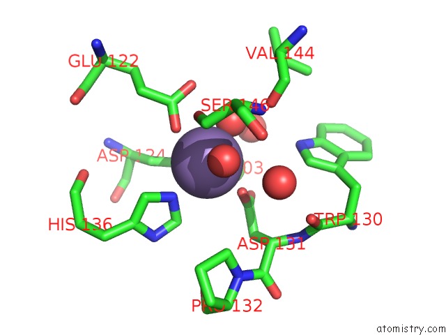
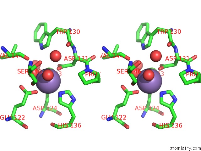
Manganese binding site 2 out of 4 in 2e7t
Go back to
Manganese binding site 2 out
of 4 in the Crystal Structure of Basic Winged Bean Lectin in Complex with A Blood Group Trisaccharide
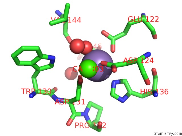
Mono view
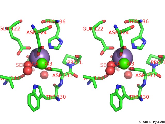
Stereo pair view
Mono view
Stereo pair view
A full contact list of Manganese with other atoms in the Mn binding
site number 2 of Crystal Structure of Basic Winged Bean Lectin in Complex with A Blood Group Trisaccharide within 5.0Å range:
|
Manganese binding site 3 out of 4 in 2e7t
Go back to
Manganese binding site 3 out
of 4 in the Crystal Structure of Basic Winged Bean Lectin in Complex with A Blood Group Trisaccharide
Mono view
Stereo pair view
Mono view
Stereo pair view
A full contact list of Manganese with other atoms in the Mn binding
site number 3 of Crystal Structure of Basic Winged Bean Lectin in Complex with A Blood Group Trisaccharide within 5.0Å range:
|
Manganese binding site 4 out of 4 in 2e7t
Go back to
Manganese binding site 4 out
of 4 in the Crystal Structure of Basic Winged Bean Lectin in Complex with A Blood Group Trisaccharide
Mono view
Stereo pair view
Mono view
Stereo pair view
A full contact list of Manganese with other atoms in the Mn binding
site number 4 of Crystal Structure of Basic Winged Bean Lectin in Complex with A Blood Group Trisaccharide within 5.0Å range:
|
Reference:
K.A.Kulkarni,
S.Katiyar,
A.Surolia,
M.Vijayan,
K.Suguna.
Generation of Blood Group Specificity: New Insights From Structural Studies on the Complexes of A- and B-Reactive Saccharides with Basic Winged Bean Agglutinin Proteins V. 68 762 2007.
ISSN: ISSN 0887-3585
PubMed: 17510954
DOI: 10.1002/PROT.21428
Page generated: Sat Oct 5 13:57:40 2024
ISSN: ISSN 0887-3585
PubMed: 17510954
DOI: 10.1002/PROT.21428
Last articles
Zn in 9MJ5Zn in 9HNW
Zn in 9G0L
Zn in 9FNE
Zn in 9DZN
Zn in 9E0I
Zn in 9D32
Zn in 9DAK
Zn in 8ZXC
Zn in 8ZUF