Manganese »
PDB 2dvd-2fer »
2dvd »
Manganese in PDB 2dvd: Crystal Structure of Peanut Lectin Gal-Alpha-1,3-Gal Complex
Protein crystallography data
The structure of Crystal Structure of Peanut Lectin Gal-Alpha-1,3-Gal Complex, PDB code: 2dvd
was solved by
S.K.Natchiar,
O.Srinivas,
N.Mitra,
A.Surolia,
N.Jayaraman,
M.Vijayan,
with X-Ray Crystallography technique. A brief refinement statistics is given in the table below:
| Resolution Low / High (Å) | 19.96 / 2.25 |
| Space group | P 21 21 2 |
| Cell size a, b, c (Å), α, β, γ (°) | 126.440, 124.640, 75.690, 90.00, 90.00, 90.00 |
| R / Rfree (%) | 18.4 / 23.2 |
Other elements in 2dvd:
The structure of Crystal Structure of Peanut Lectin Gal-Alpha-1,3-Gal Complex also contains other interesting chemical elements:
| Calcium | (Ca) | 4 atoms |
Manganese Binding Sites:
The binding sites of Manganese atom in the Crystal Structure of Peanut Lectin Gal-Alpha-1,3-Gal Complex
(pdb code 2dvd). This binding sites where shown within
5.0 Angstroms radius around Manganese atom.
In total 4 binding sites of Manganese where determined in the Crystal Structure of Peanut Lectin Gal-Alpha-1,3-Gal Complex, PDB code: 2dvd:
Jump to Manganese binding site number: 1; 2; 3; 4;
In total 4 binding sites of Manganese where determined in the Crystal Structure of Peanut Lectin Gal-Alpha-1,3-Gal Complex, PDB code: 2dvd:
Jump to Manganese binding site number: 1; 2; 3; 4;
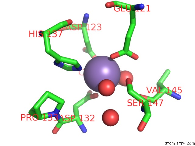
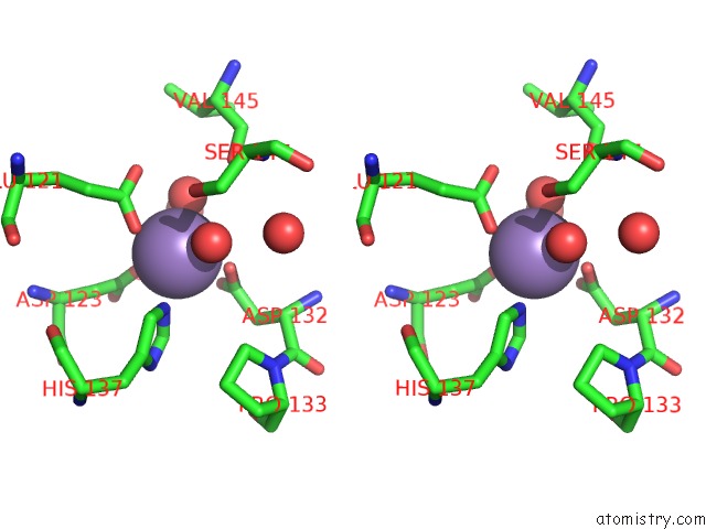
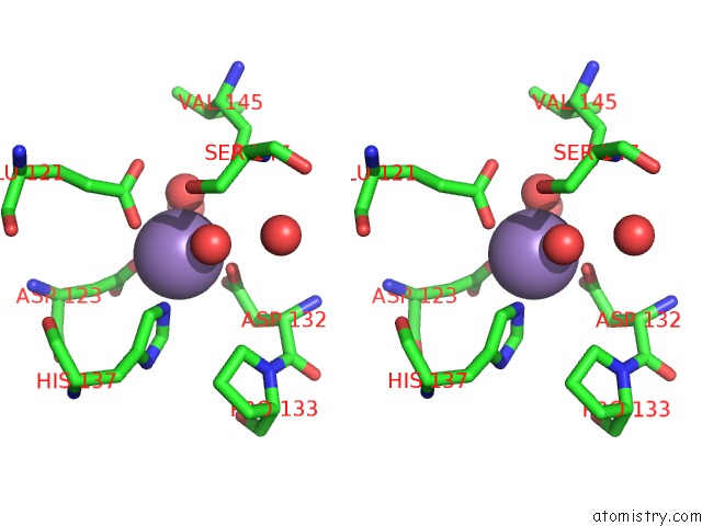
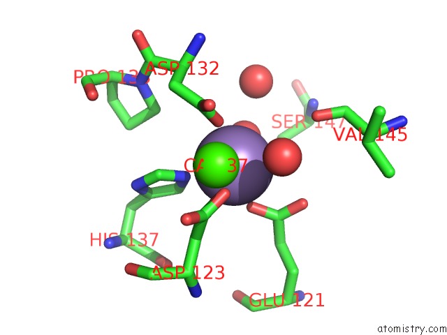
Manganese binding site 1 out of 4 in 2dvd
Go back to
Manganese binding site 1 out
of 4 in the Crystal Structure of Peanut Lectin Gal-Alpha-1,3-Gal Complex
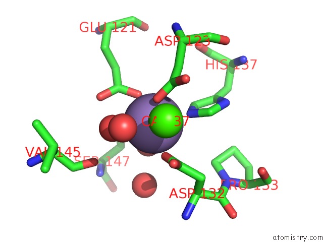
Mono view
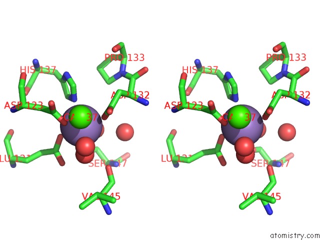
Stereo pair view
Mono view
Stereo pair view
A full contact list of Manganese with other atoms in the Mn binding
site number 1 of Crystal Structure of Peanut Lectin Gal-Alpha-1,3-Gal Complex within 5.0Å range:
|
Manganese binding site 2 out of 4 in 2dvd
Go back to
Manganese binding site 2 out
of 4 in the Crystal Structure of Peanut Lectin Gal-Alpha-1,3-Gal Complex
Mono view
Stereo pair view
Mono view
Stereo pair view
A full contact list of Manganese with other atoms in the Mn binding
site number 2 of Crystal Structure of Peanut Lectin Gal-Alpha-1,3-Gal Complex within 5.0Å range:
|
Manganese binding site 3 out of 4 in 2dvd
Go back to
Manganese binding site 3 out
of 4 in the Crystal Structure of Peanut Lectin Gal-Alpha-1,3-Gal Complex

Mono view
Stereo pair view
Mono view
Stereo pair view
A full contact list of Manganese with other atoms in the Mn binding
site number 3 of Crystal Structure of Peanut Lectin Gal-Alpha-1,3-Gal Complex within 5.0Å range:
|
Manganese binding site 4 out of 4 in 2dvd
Go back to
Manganese binding site 4 out
of 4 in the Crystal Structure of Peanut Lectin Gal-Alpha-1,3-Gal Complex
Mono view

Stereo pair view
Mono view
Stereo pair view
A full contact list of Manganese with other atoms in the Mn binding
site number 4 of Crystal Structure of Peanut Lectin Gal-Alpha-1,3-Gal Complex within 5.0Å range:
|
Reference:
S.K.Natchiar,
O.Srinivas,
N.Mitra,
A.Surolia,
N.Jayaraman,
M.Vijayan.
Structural Studies on Peanut Lectin Complexed with Disaccharides Involving Different Linkages: Further Insights Into the Structure and Interactions of the Lectin Acta Crystallogr.,Sect.D V. 62 1413 2006.
ISSN: ISSN 0907-4449
PubMed: 17057347
DOI: 10.1107/S0907444906035712
Page generated: Sat Oct 5 13:55:15 2024
ISSN: ISSN 0907-4449
PubMed: 17057347
DOI: 10.1107/S0907444906035712
Last articles
Fe in 2YXOFe in 2YRS
Fe in 2YXC
Fe in 2YNM
Fe in 2YVJ
Fe in 2YP1
Fe in 2YU2
Fe in 2YU1
Fe in 2YQB
Fe in 2YOO