Manganese »
PDB 2cev-2dvb »
2dfj »
Manganese in PDB 2dfj: Crystal Structure of the Diadenosine Tetraphosphate Hydrolase From Shigella Flexneri 2A
Enzymatic activity of Crystal Structure of the Diadenosine Tetraphosphate Hydrolase From Shigella Flexneri 2A
All present enzymatic activity of Crystal Structure of the Diadenosine Tetraphosphate Hydrolase From Shigella Flexneri 2A:
3.6.1.41;
3.6.1.41;
Protein crystallography data
The structure of Crystal Structure of the Diadenosine Tetraphosphate Hydrolase From Shigella Flexneri 2A, PDB code: 2dfj
was solved by
Q.H.Wang,
W.X.Hu,
R.C.Bi,
with X-Ray Crystallography technique. A brief refinement statistics is given in the table below:
| Resolution Low / High (Å) | 38.95 / 2.72 |
| Space group | C 1 2 1 |
| Cell size a, b, c (Å), α, β, γ (°) | 167.498, 54.965, 119.203, 90.00, 129.14, 90.00 |
| R / Rfree (%) | 22.9 / 26.8 |
Manganese Binding Sites:
The binding sites of Manganese atom in the Crystal Structure of the Diadenosine Tetraphosphate Hydrolase From Shigella Flexneri 2A
(pdb code 2dfj). This binding sites where shown within
5.0 Angstroms radius around Manganese atom.
In total 4 binding sites of Manganese where determined in the Crystal Structure of the Diadenosine Tetraphosphate Hydrolase From Shigella Flexneri 2A, PDB code: 2dfj:
Jump to Manganese binding site number: 1; 2; 3; 4;
In total 4 binding sites of Manganese where determined in the Crystal Structure of the Diadenosine Tetraphosphate Hydrolase From Shigella Flexneri 2A, PDB code: 2dfj:
Jump to Manganese binding site number: 1; 2; 3; 4;
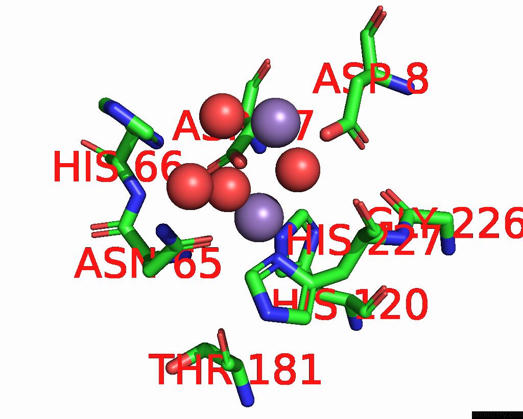
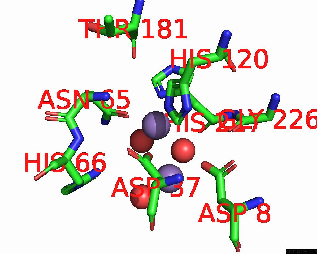
Manganese binding site 1 out of 4 in 2dfj
Go back to
Manganese binding site 1 out
of 4 in the Crystal Structure of the Diadenosine Tetraphosphate Hydrolase From Shigella Flexneri 2A
Mono view
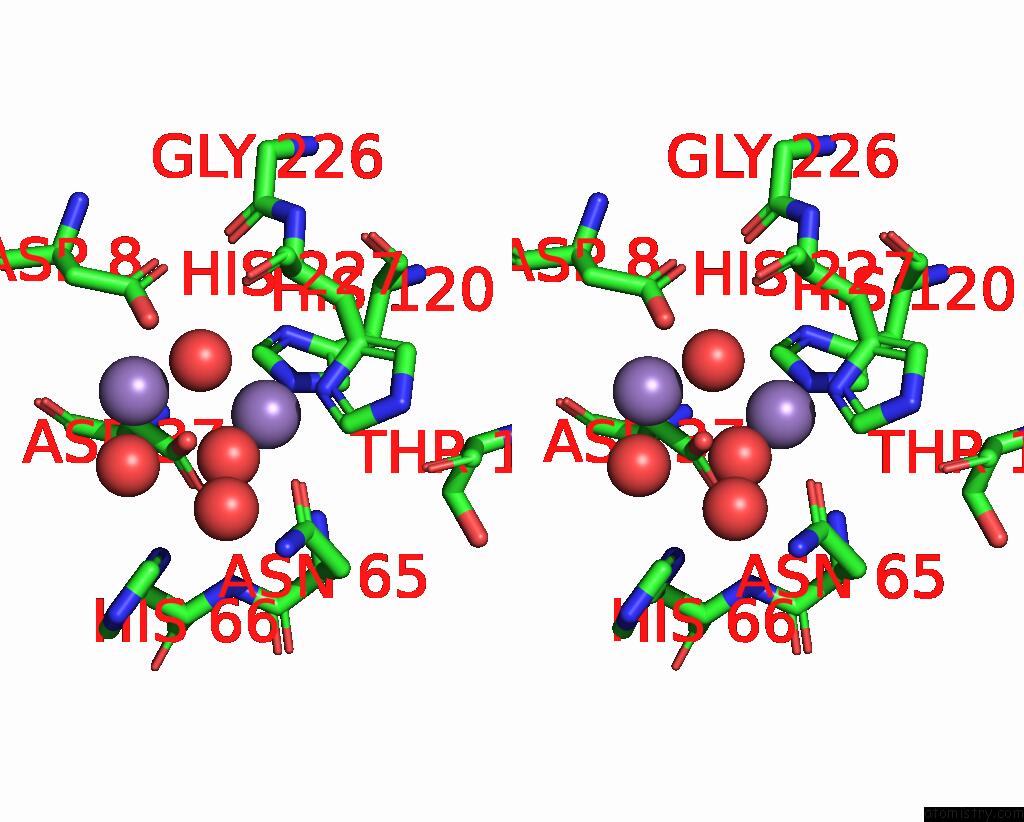
Stereo pair view
Mono view
Stereo pair view
A full contact list of Manganese with other atoms in the Mn binding
site number 1 of Crystal Structure of the Diadenosine Tetraphosphate Hydrolase From Shigella Flexneri 2A within 5.0Å range:
|
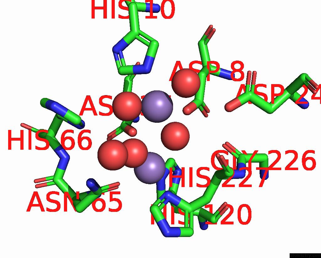
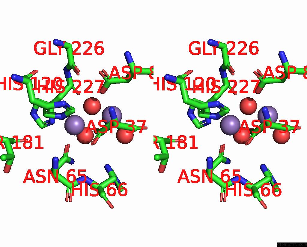
Manganese binding site 2 out of 4 in 2dfj
Go back to
Manganese binding site 2 out
of 4 in the Crystal Structure of the Diadenosine Tetraphosphate Hydrolase From Shigella Flexneri 2A
Mono view
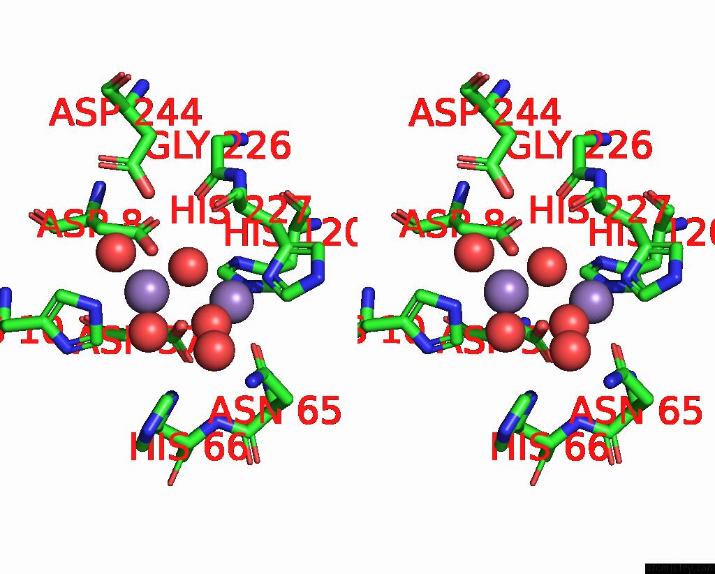
Stereo pair view
Mono view
Stereo pair view
A full contact list of Manganese with other atoms in the Mn binding
site number 2 of Crystal Structure of the Diadenosine Tetraphosphate Hydrolase From Shigella Flexneri 2A within 5.0Å range:
|
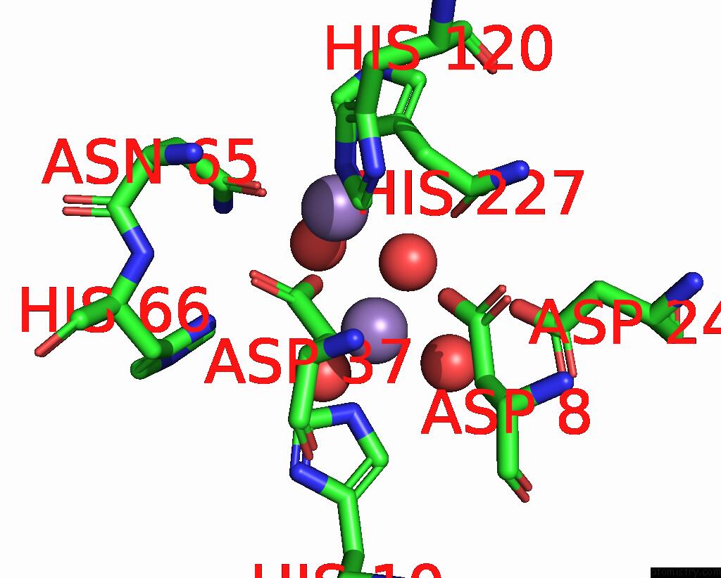
Manganese binding site 3 out of 4 in 2dfj
Go back to
Manganese binding site 3 out
of 4 in the Crystal Structure of the Diadenosine Tetraphosphate Hydrolase From Shigella Flexneri 2A
Mono view
Stereo pair view
Mono view
Stereo pair view
A full contact list of Manganese with other atoms in the Mn binding
site number 3 of Crystal Structure of the Diadenosine Tetraphosphate Hydrolase From Shigella Flexneri 2A within 5.0Å range:
|
Manganese binding site 4 out of 4 in 2dfj
Go back to
Manganese binding site 4 out
of 4 in the Crystal Structure of the Diadenosine Tetraphosphate Hydrolase From Shigella Flexneri 2A
Mono view
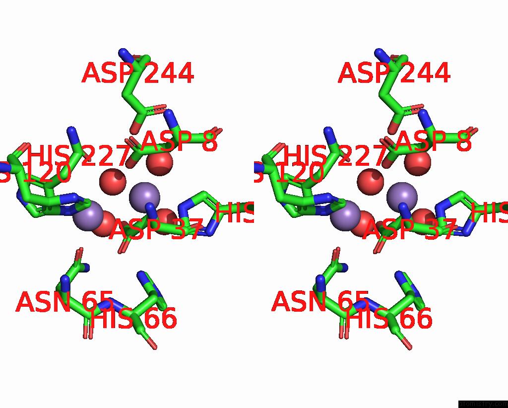
Stereo pair view
Mono view
Stereo pair view
A full contact list of Manganese with other atoms in the Mn binding
site number 4 of Crystal Structure of the Diadenosine Tetraphosphate Hydrolase From Shigella Flexneri 2A within 5.0Å range:
|
Reference:
Q.H.Wang,
W.X.Hu,
W.Gao,
R.C.Bi.
Crystal Structure of the Diadenosine Tetraphosphate Hydrolase From Shigella Flexneri 2A Proteins V. 65 1032 2006.
ISSN: ISSN 0887-3585
PubMed: 17006950
DOI: 10.1002/PROT.21106
Page generated: Sat Oct 5 13:45:26 2024
ISSN: ISSN 0887-3585
PubMed: 17006950
DOI: 10.1002/PROT.21106
Last articles
Zn in 9MJ5Zn in 9HNW
Zn in 9G0L
Zn in 9FNE
Zn in 9DZN
Zn in 9E0I
Zn in 9D32
Zn in 9DAK
Zn in 8ZXC
Zn in 8ZUF