Manganese »
PDB 2cev-2dvb »
2d7r »
Manganese in PDB 2d7r: Crystal Structure of Pp-Galnac-T10 Complexed with Galnac-Ser on Lectin Domain
Enzymatic activity of Crystal Structure of Pp-Galnac-T10 Complexed with Galnac-Ser on Lectin Domain
All present enzymatic activity of Crystal Structure of Pp-Galnac-T10 Complexed with Galnac-Ser on Lectin Domain:
2.4.1.41;
2.4.1.41;
Protein crystallography data
The structure of Crystal Structure of Pp-Galnac-T10 Complexed with Galnac-Ser on Lectin Domain, PDB code: 2d7r
was solved by
T.Kubota,
T.Shiba,
S.Sugioka,
R.Kato,
S.Wakatsuki,
H.Narimatsu,
with X-Ray Crystallography technique. A brief refinement statistics is given in the table below:
| Resolution Low / High (Å) | 40.00 / 2.80 |
| Space group | C 2 2 21 |
| Cell size a, b, c (Å), α, β, γ (°) | 80.195, 132.812, 138.953, 90.00, 90.00, 90.00 |
| R / Rfree (%) | 22.9 / 29.8 |
Manganese Binding Sites:
The binding sites of Manganese atom in the Crystal Structure of Pp-Galnac-T10 Complexed with Galnac-Ser on Lectin Domain
(pdb code 2d7r). This binding sites where shown within
5.0 Angstroms radius around Manganese atom.
In total only one binding site of Manganese was determined in the Crystal Structure of Pp-Galnac-T10 Complexed with Galnac-Ser on Lectin Domain, PDB code: 2d7r:
In total only one binding site of Manganese was determined in the Crystal Structure of Pp-Galnac-T10 Complexed with Galnac-Ser on Lectin Domain, PDB code: 2d7r:
Manganese binding site 1 out of 1 in 2d7r
Go back to
Manganese binding site 1 out
of 1 in the Crystal Structure of Pp-Galnac-T10 Complexed with Galnac-Ser on Lectin Domain
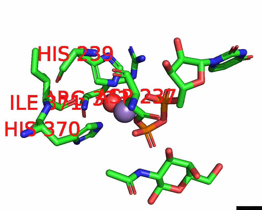
Mono view
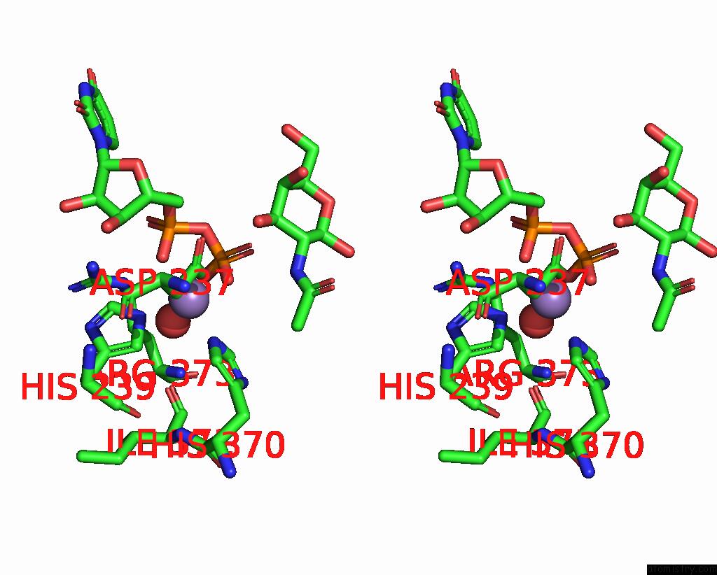
Stereo pair view
Mono view
Stereo pair view
A full contact list of Manganese with other atoms in the Mn binding
site number 1 of Crystal Structure of Pp-Galnac-T10 Complexed with Galnac-Ser on Lectin Domain within 5.0Å range:
|
Reference:
T.Kubota,
T.Shiba,
S.Sugioka,
S.Furukawa,
H.Sawaki,
R.Kato,
S.Wakatsuki,
H.Narimatsu.
Structural Basis of Carbohydrate Transfer Activity By Human Udp-Galnac: Polypeptide Alpha-N-Acetylgalactosaminyltransferase (Pp-Galnac-T10) J.Mol.Biol. V. 359 708 2006.
ISSN: ISSN 0022-2836
PubMed: 16650853
DOI: 10.1016/J.JMB.2006.03.061
Page generated: Sat Oct 5 13:44:42 2024
ISSN: ISSN 0022-2836
PubMed: 16650853
DOI: 10.1016/J.JMB.2006.03.061
Last articles
Zn in 9MJ5Zn in 9HNW
Zn in 9G0L
Zn in 9FNE
Zn in 9DZN
Zn in 9E0I
Zn in 9D32
Zn in 9DAK
Zn in 8ZXC
Zn in 8ZUF