Manganese »
PDB 2cev-2dvb »
2d0c »
Manganese in PDB 2d0c: Crystal Structure of Bst-Rnase Hiii in Complex with MN2+
Enzymatic activity of Crystal Structure of Bst-Rnase Hiii in Complex with MN2+
All present enzymatic activity of Crystal Structure of Bst-Rnase Hiii in Complex with MN2+:
3.1.26.4;
3.1.26.4;
Protein crystallography data
The structure of Crystal Structure of Bst-Rnase Hiii in Complex with MN2+, PDB code: 2d0c
was solved by
H.Chon,
H.Matsumura,
Y.Koga,
K.Takano,
S.Kanaya,
with X-Ray Crystallography technique. A brief refinement statistics is given in the table below:
| Resolution Low / High (Å) | 19.64 / 2.60 |
| Space group | P 21 21 2 |
| Cell size a, b, c (Å), α, β, γ (°) | 67.288, 109.771, 48.370, 90.00, 90.00, 90.00 |
| R / Rfree (%) | 20.9 / 27.4 |
Manganese Binding Sites:
The binding sites of Manganese atom in the Crystal Structure of Bst-Rnase Hiii in Complex with MN2+
(pdb code 2d0c). This binding sites where shown within
5.0 Angstroms radius around Manganese atom.
In total only one binding site of Manganese was determined in the Crystal Structure of Bst-Rnase Hiii in Complex with MN2+, PDB code: 2d0c:
In total only one binding site of Manganese was determined in the Crystal Structure of Bst-Rnase Hiii in Complex with MN2+, PDB code: 2d0c:
Manganese binding site 1 out of 1 in 2d0c
Go back to
Manganese binding site 1 out
of 1 in the Crystal Structure of Bst-Rnase Hiii in Complex with MN2+
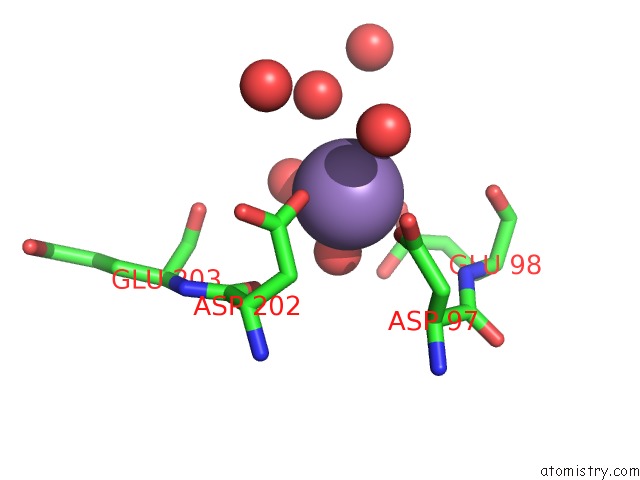
Mono view
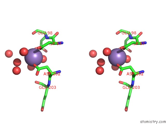
Stereo pair view
Mono view
Stereo pair view
A full contact list of Manganese with other atoms in the Mn binding
site number 1 of Crystal Structure of Bst-Rnase Hiii in Complex with MN2+ within 5.0Å range:
|
Reference:
H.Chon,
H.Matsumura,
Y.Koga,
K.Takano,
S.Kanaya.
Crystal Structure and Structure-Based Mutational Analyses of Rnase Hiii From Bacillus Stearothermophilus: A New Type 2 Rnase H with Tbp-Like Substrate-Binding Domain at the N Terminus J.Mol.Biol. V. 356 165 2006.
ISSN: ISSN 0022-2836
PubMed: 16343535
DOI: 10.1016/J.JMB.2005.11.017
Page generated: Sat Oct 5 13:40:11 2024
ISSN: ISSN 0022-2836
PubMed: 16343535
DOI: 10.1016/J.JMB.2005.11.017
Last articles
Fe in 2YXOFe in 2YRS
Fe in 2YXC
Fe in 2YNM
Fe in 2YVJ
Fe in 2YP1
Fe in 2YU2
Fe in 2YU1
Fe in 2YQB
Fe in 2YOO