Manganese »
PDB 2ayl-2ce4 »
2bqp »
Manganese in PDB 2bqp: The Structure of the Pea Lectin-D-Glucopyranose Complex
Protein crystallography data
The structure of The Structure of the Pea Lectin-D-Glucopyranose Complex, PDB code: 2bqp
was solved by
S.N.Ruzeinikov,
I.Mikhailova Yu,
I.N.Tsygannik,
W.Pangborn,
W.Duax,
V.Z.Pletnev,
with X-Ray Crystallography technique. A brief refinement statistics is given in the table below:
| Resolution Low / High (Å) | 8.00 / 1.90 |
| Space group | P 21 21 21 |
| Cell size a, b, c (Å), α, β, γ (°) | 62.838, 135.246, 54.781, 90.00, 90.00, 90.00 |
| R / Rfree (%) | 16.1 / 18.8 |
Other elements in 2bqp:
The structure of The Structure of the Pea Lectin-D-Glucopyranose Complex also contains other interesting chemical elements:
| Calcium | (Ca) | 2 atoms |
Manganese Binding Sites:
The binding sites of Manganese atom in the The Structure of the Pea Lectin-D-Glucopyranose Complex
(pdb code 2bqp). This binding sites where shown within
5.0 Angstroms radius around Manganese atom.
In total 2 binding sites of Manganese where determined in the The Structure of the Pea Lectin-D-Glucopyranose Complex, PDB code: 2bqp:
Jump to Manganese binding site number: 1; 2;
In total 2 binding sites of Manganese where determined in the The Structure of the Pea Lectin-D-Glucopyranose Complex, PDB code: 2bqp:
Jump to Manganese binding site number: 1; 2;
Manganese binding site 1 out of 2 in 2bqp
Go back to
Manganese binding site 1 out
of 2 in the The Structure of the Pea Lectin-D-Glucopyranose Complex
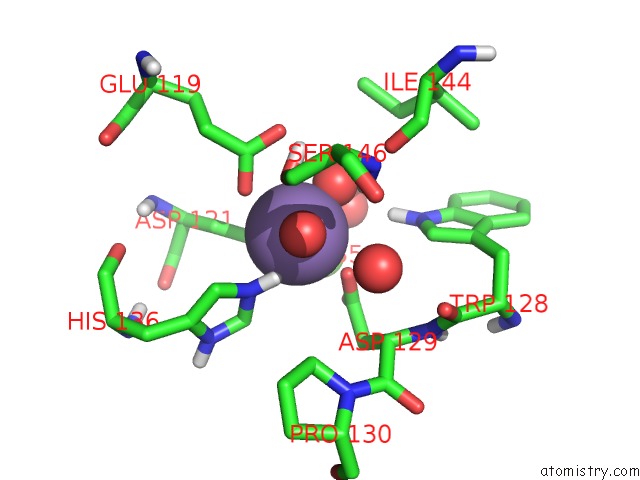
Mono view
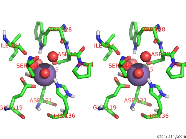
Stereo pair view
Mono view
Stereo pair view
A full contact list of Manganese with other atoms in the Mn binding
site number 1 of The Structure of the Pea Lectin-D-Glucopyranose Complex within 5.0Å range:
|
Manganese binding site 2 out of 2 in 2bqp
Go back to
Manganese binding site 2 out
of 2 in the The Structure of the Pea Lectin-D-Glucopyranose Complex
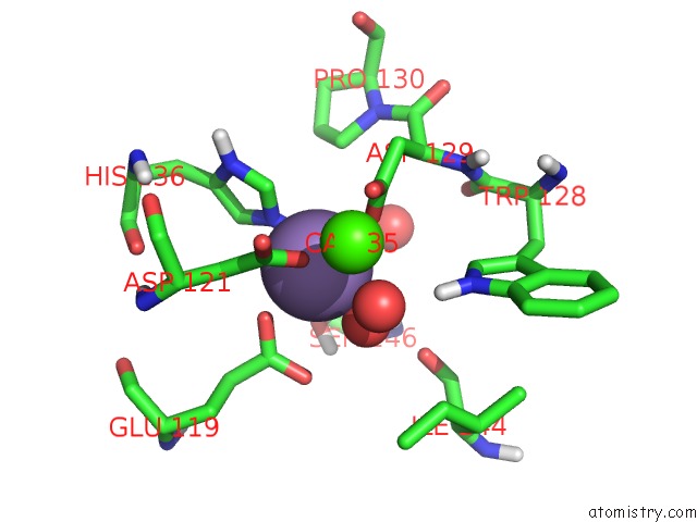
Mono view
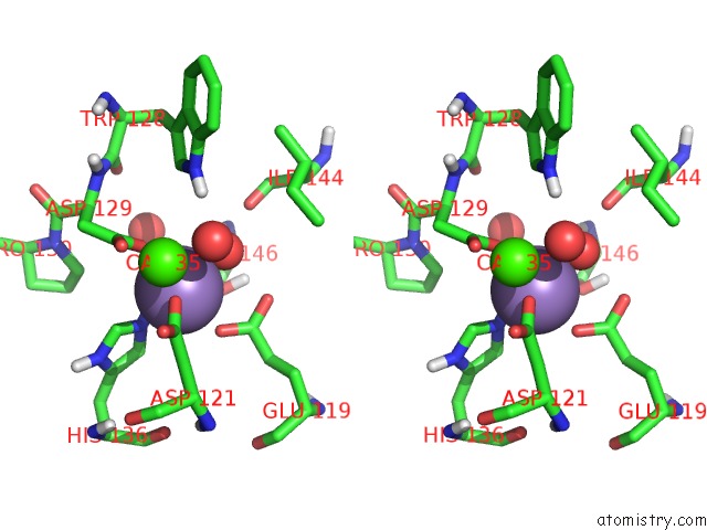
Stereo pair view
Mono view
Stereo pair view
A full contact list of Manganese with other atoms in the Mn binding
site number 2 of The Structure of the Pea Lectin-D-Glucopyranose Complex within 5.0Å range:
|
Reference:
V.Z.Pletnev,
S.N.Ruzheinikov,
I.N.Tsygannik,
I.Mikhailova Yu,
W.Duax,
D.Ghosh,
W.Pangborn.
The Structure of Pea Lectin-D-Glucopyranose Complex at A 1.9 A Resolution Russ.J.Bioorganic Chem. V. 23 469 1997.
ISSN: ISSN 1068-1620
Page generated: Sat Oct 5 13:35:23 2024
ISSN: ISSN 1068-1620
Last articles
Zn in 9JYWZn in 9IR4
Zn in 9IR3
Zn in 9GMX
Zn in 9GMW
Zn in 9JEJ
Zn in 9ERF
Zn in 9ERE
Zn in 9EGV
Zn in 9EGW