Manganese »
PDB 2ayl-2ce4 »
2bgg »
Manganese in PDB 2bgg: The Structure of A Piwi Protein From Archaeoglobus Fulgidus Complexed with A 16NT Sirna Duplex.
Protein crystallography data
The structure of The Structure of A Piwi Protein From Archaeoglobus Fulgidus Complexed with A 16NT Sirna Duplex., PDB code: 2bgg
was solved by
J.S.Parker,
S.M.Roe,
D.Barford,
with X-Ray Crystallography technique. A brief refinement statistics is given in the table below:
| Resolution Low / High (Å) | 99.01 / 2.20 |
| Space group | P 1 |
| Cell size a, b, c (Å), α, β, γ (°) | 51.916, 61.206, 104.059, 76.54, 76.14, 79.35 |
| R / Rfree (%) | 18 / 24.3 |
Manganese Binding Sites:
The binding sites of Manganese atom in the The Structure of A Piwi Protein From Archaeoglobus Fulgidus Complexed with A 16NT Sirna Duplex.
(pdb code 2bgg). This binding sites where shown within
5.0 Angstroms radius around Manganese atom.
In total 2 binding sites of Manganese where determined in the The Structure of A Piwi Protein From Archaeoglobus Fulgidus Complexed with A 16NT Sirna Duplex., PDB code: 2bgg:
Jump to Manganese binding site number: 1; 2;
In total 2 binding sites of Manganese where determined in the The Structure of A Piwi Protein From Archaeoglobus Fulgidus Complexed with A 16NT Sirna Duplex., PDB code: 2bgg:
Jump to Manganese binding site number: 1; 2;
Manganese binding site 1 out of 2 in 2bgg
Go back to
Manganese binding site 1 out
of 2 in the The Structure of A Piwi Protein From Archaeoglobus Fulgidus Complexed with A 16NT Sirna Duplex.
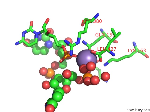
Mono view
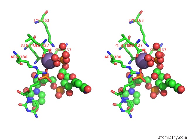
Stereo pair view
Mono view
Stereo pair view
A full contact list of Manganese with other atoms in the Mn binding
site number 1 of The Structure of A Piwi Protein From Archaeoglobus Fulgidus Complexed with A 16NT Sirna Duplex. within 5.0Å range:
|
Manganese binding site 2 out of 2 in 2bgg
Go back to
Manganese binding site 2 out
of 2 in the The Structure of A Piwi Protein From Archaeoglobus Fulgidus Complexed with A 16NT Sirna Duplex.
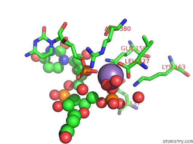
Mono view
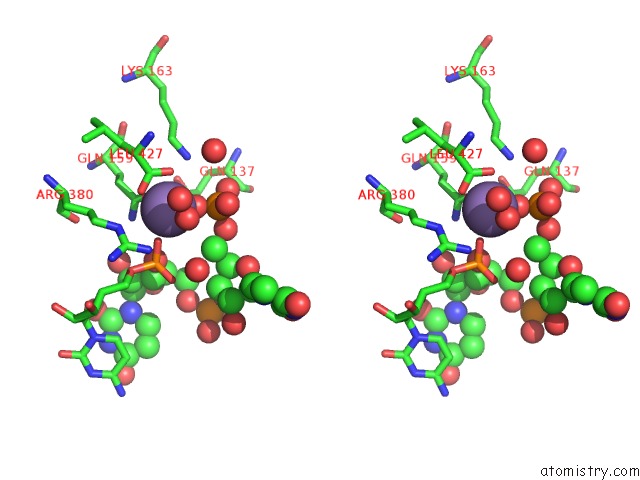
Stereo pair view
Mono view
Stereo pair view
A full contact list of Manganese with other atoms in the Mn binding
site number 2 of The Structure of A Piwi Protein From Archaeoglobus Fulgidus Complexed with A 16NT Sirna Duplex. within 5.0Å range:
|
Reference:
J.S.Parker,
S.M.Roe,
D.Barford.
Structural Insights Into Mrna Recognition From A Piwi Domain-Sirna Guide Complex Nature V. 434 663 2005.
ISSN: ISSN 0028-0836
PubMed: 15800628
DOI: 10.1038/NATURE03462
Page generated: Sat Oct 5 13:34:25 2024
ISSN: ISSN 0028-0836
PubMed: 15800628
DOI: 10.1038/NATURE03462
Last articles
Ca in 5SZMCa in 5SZL
Ca in 5SY1
Ca in 5SWI
Ca in 5SVE
Ca in 5SSX
Ca in 5SV0
Ca in 5STD
Ca in 5SSZ
Ca in 5SSY