Manganese »
PDB 2ayl-2ce4 »
2bcd »
Manganese in PDB 2bcd: X-Ray Crystal Structure of Protein Phosphatase-1 with the Marine Toxin Motuporin Bound
Enzymatic activity of X-Ray Crystal Structure of Protein Phosphatase-1 with the Marine Toxin Motuporin Bound
All present enzymatic activity of X-Ray Crystal Structure of Protein Phosphatase-1 with the Marine Toxin Motuporin Bound:
3.1.3.16;
3.1.3.16;
Protein crystallography data
The structure of X-Ray Crystal Structure of Protein Phosphatase-1 with the Marine Toxin Motuporin Bound, PDB code: 2bcd
was solved by
J.T.Maynes,
H.A.Luu,
M.M.Cherney,
R.J.Andersen,
D.Williams,
C.F.Holmes,
M.N.James,
with X-Ray Crystallography technique. A brief refinement statistics is given in the table below:
| Resolution Low / High (Å) | 31.75 / 2.10 |
| Space group | P 42 21 2 |
| Cell size a, b, c (Å), α, β, γ (°) | 100.955, 100.955, 63.485, 90.00, 90.00, 90.00 |
| R / Rfree (%) | 22.1 / 26.4 |
Manganese Binding Sites:
The binding sites of Manganese atom in the X-Ray Crystal Structure of Protein Phosphatase-1 with the Marine Toxin Motuporin Bound
(pdb code 2bcd). This binding sites where shown within
5.0 Angstroms radius around Manganese atom.
In total 8 binding sites of Manganese where determined in the X-Ray Crystal Structure of Protein Phosphatase-1 with the Marine Toxin Motuporin Bound, PDB code: 2bcd:
Jump to Manganese binding site number: 1; 2; 3; 4; 5; 6; 7; 8;
In total 8 binding sites of Manganese where determined in the X-Ray Crystal Structure of Protein Phosphatase-1 with the Marine Toxin Motuporin Bound, PDB code: 2bcd:
Jump to Manganese binding site number: 1; 2; 3; 4; 5; 6; 7; 8;
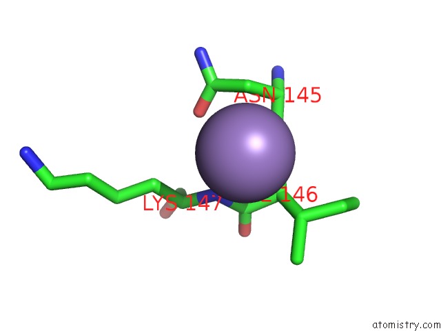
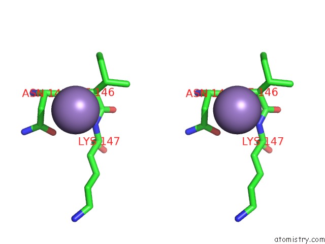
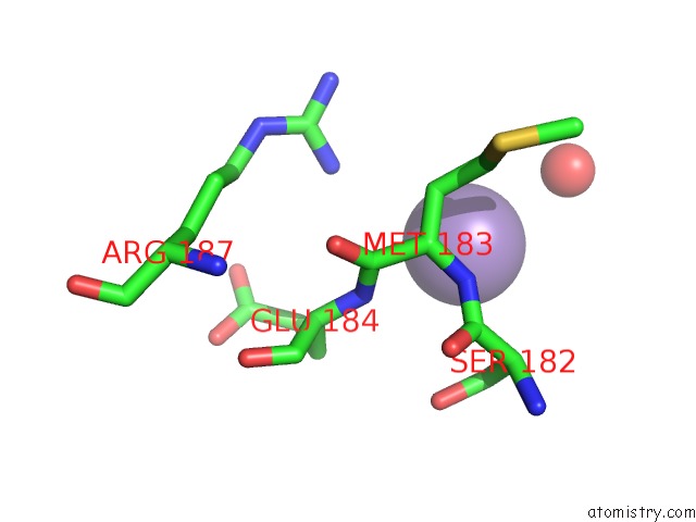
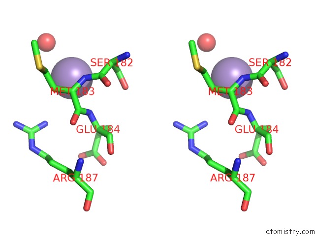
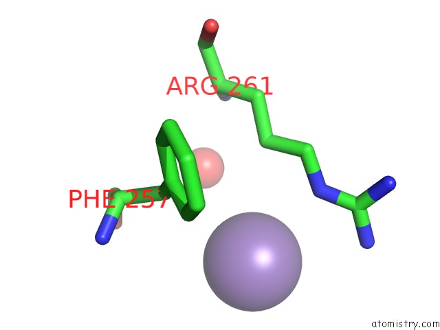
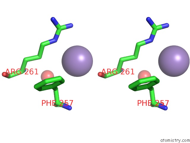
Manganese binding site 1 out of 8 in 2bcd
Go back to
Manganese binding site 1 out
of 8 in the X-Ray Crystal Structure of Protein Phosphatase-1 with the Marine Toxin Motuporin Bound
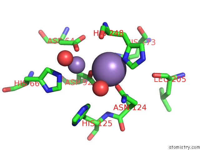
Mono view
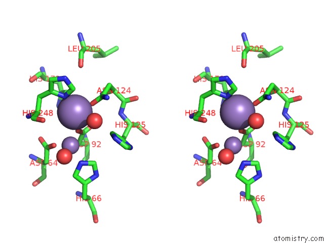
Stereo pair view
Mono view
Stereo pair view
A full contact list of Manganese with other atoms in the Mn binding
site number 1 of X-Ray Crystal Structure of Protein Phosphatase-1 with the Marine Toxin Motuporin Bound within 5.0Å range:
|
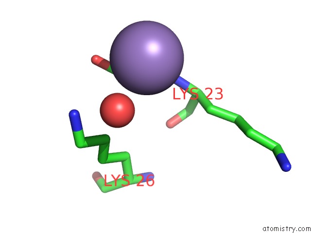
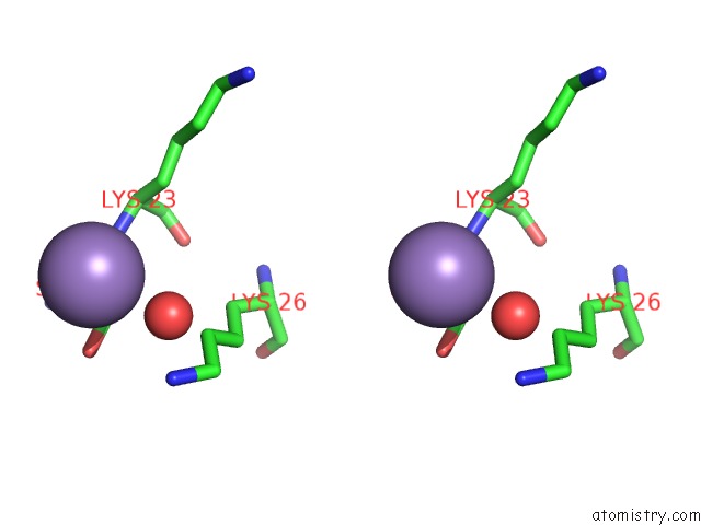
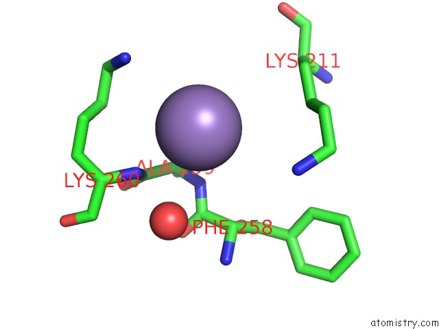
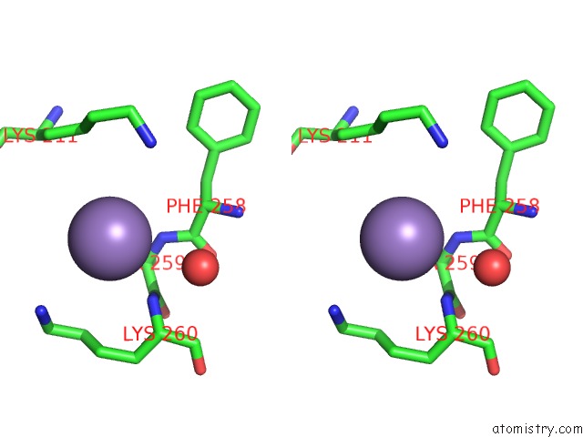
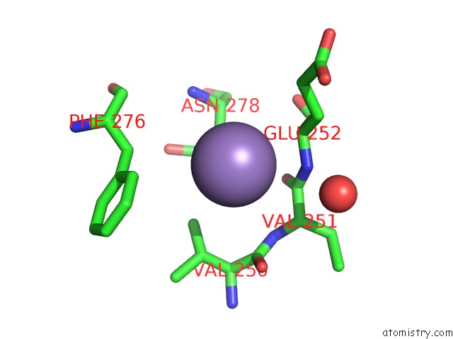
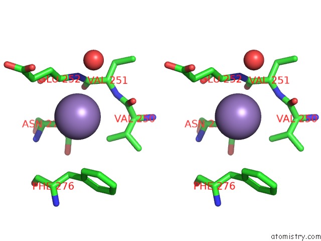
Manganese binding site 2 out of 8 in 2bcd
Go back to
Manganese binding site 2 out
of 8 in the X-Ray Crystal Structure of Protein Phosphatase-1 with the Marine Toxin Motuporin Bound
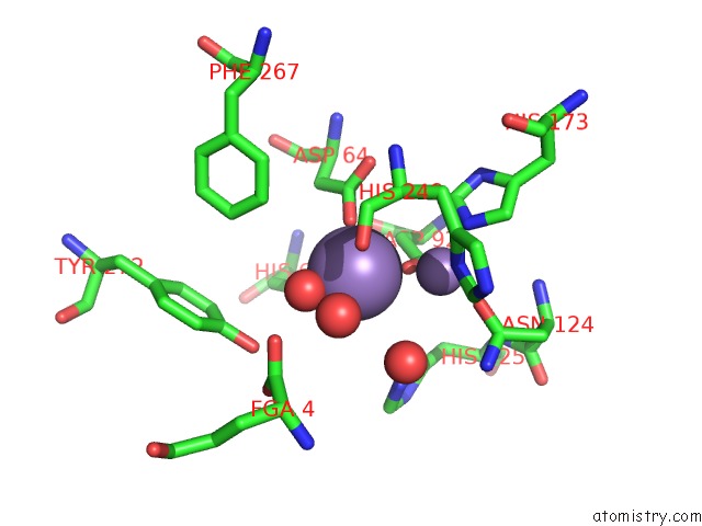
Mono view
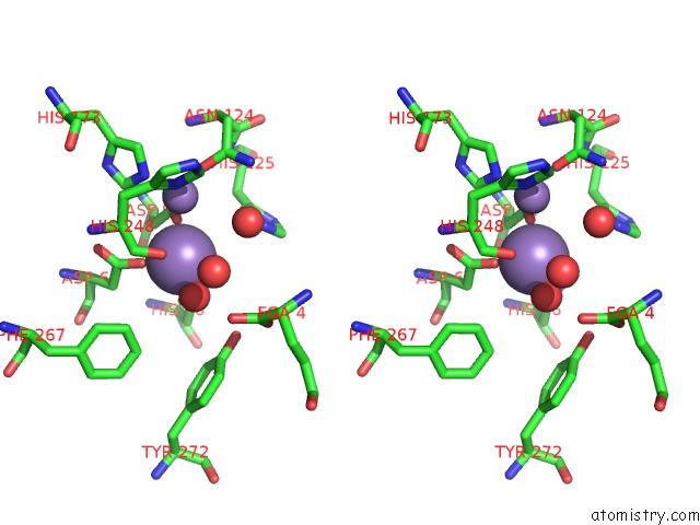
Stereo pair view
Mono view
Stereo pair view
A full contact list of Manganese with other atoms in the Mn binding
site number 2 of X-Ray Crystal Structure of Protein Phosphatase-1 with the Marine Toxin Motuporin Bound within 5.0Å range:
|
Manganese binding site 3 out of 8 in 2bcd
Go back to
Manganese binding site 3 out
of 8 in the X-Ray Crystal Structure of Protein Phosphatase-1 with the Marine Toxin Motuporin Bound
Mono view
Stereo pair view
Mono view
Stereo pair view
A full contact list of Manganese with other atoms in the Mn binding
site number 3 of X-Ray Crystal Structure of Protein Phosphatase-1 with the Marine Toxin Motuporin Bound within 5.0Å range:
|
Manganese binding site 4 out of 8 in 2bcd
Go back to
Manganese binding site 4 out
of 8 in the X-Ray Crystal Structure of Protein Phosphatase-1 with the Marine Toxin Motuporin Bound
Mono view
Stereo pair view
Mono view
Stereo pair view
A full contact list of Manganese with other atoms in the Mn binding
site number 4 of X-Ray Crystal Structure of Protein Phosphatase-1 with the Marine Toxin Motuporin Bound within 5.0Å range:
|
Manganese binding site 5 out of 8 in 2bcd
Go back to
Manganese binding site 5 out
of 8 in the X-Ray Crystal Structure of Protein Phosphatase-1 with the Marine Toxin Motuporin Bound
Mono view
Stereo pair view
Mono view
Stereo pair view
A full contact list of Manganese with other atoms in the Mn binding
site number 5 of X-Ray Crystal Structure of Protein Phosphatase-1 with the Marine Toxin Motuporin Bound within 5.0Å range:
|
Manganese binding site 6 out of 8 in 2bcd
Go back to
Manganese binding site 6 out
of 8 in the X-Ray Crystal Structure of Protein Phosphatase-1 with the Marine Toxin Motuporin Bound
Mono view
Stereo pair view
Mono view
Stereo pair view
A full contact list of Manganese with other atoms in the Mn binding
site number 6 of X-Ray Crystal Structure of Protein Phosphatase-1 with the Marine Toxin Motuporin Bound within 5.0Å range:
|
Manganese binding site 7 out of 8 in 2bcd
Go back to
Manganese binding site 7 out
of 8 in the X-Ray Crystal Structure of Protein Phosphatase-1 with the Marine Toxin Motuporin Bound
Mono view
Stereo pair view
Mono view
Stereo pair view
A full contact list of Manganese with other atoms in the Mn binding
site number 7 of X-Ray Crystal Structure of Protein Phosphatase-1 with the Marine Toxin Motuporin Bound within 5.0Å range:
|
Manganese binding site 8 out of 8 in 2bcd
Go back to
Manganese binding site 8 out
of 8 in the X-Ray Crystal Structure of Protein Phosphatase-1 with the Marine Toxin Motuporin Bound
Mono view
Stereo pair view
Mono view
Stereo pair view
A full contact list of Manganese with other atoms in the Mn binding
site number 8 of X-Ray Crystal Structure of Protein Phosphatase-1 with the Marine Toxin Motuporin Bound within 5.0Å range:
|
Reference:
J.T.Maynes,
H.A.Luu,
M.M.Cherney,
R.J.Andersen,
D.Williams,
C.F.Holmes,
M.N.James.
Crystal Structures of Protein Phosphatase-1 Bound to Motuporin and Dihydromicrocystin-La: Elucidation of the Mechanism of Enzyme Inhibition By Cyanobacterial Toxins. J.Mol.Biol. V. 356 111 2006.
ISSN: ISSN 0022-2836
PubMed: 16343532
DOI: 10.1016/J.JMB.2005.11.019
Page generated: Sat Oct 5 13:32:54 2024
ISSN: ISSN 0022-2836
PubMed: 16343532
DOI: 10.1016/J.JMB.2005.11.019
Last articles
Zn in 9MJ5Zn in 9HNW
Zn in 9G0L
Zn in 9FNE
Zn in 9DZN
Zn in 9E0I
Zn in 9D32
Zn in 9DAK
Zn in 8ZXC
Zn in 8ZUF