Manganese »
PDB 2ayl-2ce4 »
2b7y »
Manganese in PDB 2b7y: Fava Bean Lectin-Glucose Complex
Protein crystallography data
The structure of Fava Bean Lectin-Glucose Complex, PDB code: 2b7y
was solved by
G.N.Reeke Jr.,
J.W.Becker,
with X-Ray Crystallography technique. A brief refinement statistics is given in the table below:
| Resolution Low / High (Å) | 46.18 / 3.00 |
| Space group | P 21 21 21 |
| Cell size a, b, c (Å), α, β, γ (°) | 90.000, 89.300, 67.400, 90.00, 90.00, 90.00 |
| R / Rfree (%) | 22.4 / 30 |
Other elements in 2b7y:
The structure of Fava Bean Lectin-Glucose Complex also contains other interesting chemical elements:
| Calcium | (Ca) | 2 atoms |
Manganese Binding Sites:
The binding sites of Manganese atom in the Fava Bean Lectin-Glucose Complex
(pdb code 2b7y). This binding sites where shown within
5.0 Angstroms radius around Manganese atom.
In total 2 binding sites of Manganese where determined in the Fava Bean Lectin-Glucose Complex, PDB code: 2b7y:
Jump to Manganese binding site number: 1; 2;
In total 2 binding sites of Manganese where determined in the Fava Bean Lectin-Glucose Complex, PDB code: 2b7y:
Jump to Manganese binding site number: 1; 2;
Manganese binding site 1 out of 2 in 2b7y
Go back to
Manganese binding site 1 out
of 2 in the Fava Bean Lectin-Glucose Complex
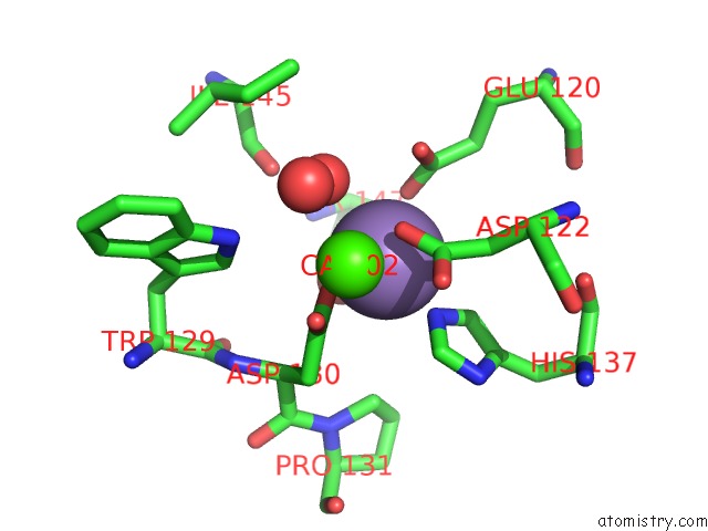
Mono view
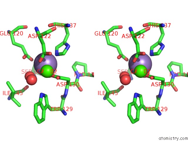
Stereo pair view
Mono view
Stereo pair view
A full contact list of Manganese with other atoms in the Mn binding
site number 1 of Fava Bean Lectin-Glucose Complex within 5.0Å range:
|
Manganese binding site 2 out of 2 in 2b7y
Go back to
Manganese binding site 2 out
of 2 in the Fava Bean Lectin-Glucose Complex
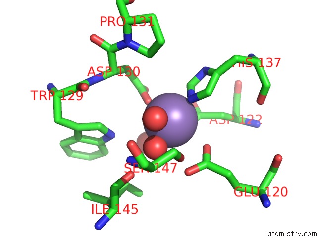
Mono view
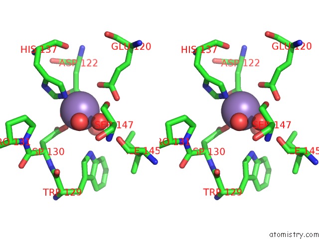
Stereo pair view
Mono view
Stereo pair view
A full contact list of Manganese with other atoms in the Mn binding
site number 2 of Fava Bean Lectin-Glucose Complex within 5.0Å range:
|
Reference:
G.N.Reeke Jr.,
J.W.Becker.
Three-Dimensional Structure of Favin: Saccharide Binding-Cyclic Permutation in Leguminous Lectins Science V. 234 1108 1986.
ISSN: ISSN 0036-8075
PubMed: 3775378
Page generated: Sat Oct 5 13:32:14 2024
ISSN: ISSN 0036-8075
PubMed: 3775378
Last articles
Zn in 9MJ5Zn in 9HNW
Zn in 9G0L
Zn in 9FNE
Zn in 9DZN
Zn in 9E0I
Zn in 9D32
Zn in 9DAK
Zn in 8ZXC
Zn in 8ZUF