Manganese »
PDB 2ayl-2ce4 »
2b7o »
Manganese in PDB 2b7o: The Structure of 3-Deoxy-D-Arabino-Heptulosonate 7-Phosphate Synthase From Mycobacterium Tuberculosis
Enzymatic activity of The Structure of 3-Deoxy-D-Arabino-Heptulosonate 7-Phosphate Synthase From Mycobacterium Tuberculosis
All present enzymatic activity of The Structure of 3-Deoxy-D-Arabino-Heptulosonate 7-Phosphate Synthase From Mycobacterium Tuberculosis:
2.5.1.54;
2.5.1.54;
Protein crystallography data
The structure of The Structure of 3-Deoxy-D-Arabino-Heptulosonate 7-Phosphate Synthase From Mycobacterium Tuberculosis, PDB code: 2b7o
was solved by
C.J.Webby,
H.M.Baker,
J.S.Lott,
E.N.Baker,
E.J.Parker,
Mycobacteriumtuberculosis Structural Proteomics Project (Xmtb),
with X-Ray Crystallography technique. A brief refinement statistics is given in the table below:
| Resolution Low / High (Å) | 47.04 / 2.30 |
| Space group | P 32 2 1 |
| Cell size a, b, c (Å), α, β, γ (°) | 204.085, 204.085, 66.230, 90.00, 90.00, 120.00 |
| R / Rfree (%) | 18.8 / 22.4 |
Manganese Binding Sites:
The binding sites of Manganese atom in the The Structure of 3-Deoxy-D-Arabino-Heptulosonate 7-Phosphate Synthase From Mycobacterium Tuberculosis
(pdb code 2b7o). This binding sites where shown within
5.0 Angstroms radius around Manganese atom.
In total 2 binding sites of Manganese where determined in the The Structure of 3-Deoxy-D-Arabino-Heptulosonate 7-Phosphate Synthase From Mycobacterium Tuberculosis, PDB code: 2b7o:
Jump to Manganese binding site number: 1; 2;
In total 2 binding sites of Manganese where determined in the The Structure of 3-Deoxy-D-Arabino-Heptulosonate 7-Phosphate Synthase From Mycobacterium Tuberculosis, PDB code: 2b7o:
Jump to Manganese binding site number: 1; 2;
Manganese binding site 1 out of 2 in 2b7o
Go back to
Manganese binding site 1 out
of 2 in the The Structure of 3-Deoxy-D-Arabino-Heptulosonate 7-Phosphate Synthase From Mycobacterium Tuberculosis
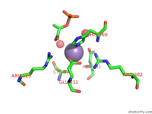
Mono view
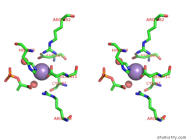
Stereo pair view
Mono view
Stereo pair view
A full contact list of Manganese with other atoms in the Mn binding
site number 1 of The Structure of 3-Deoxy-D-Arabino-Heptulosonate 7-Phosphate Synthase From Mycobacterium Tuberculosis within 5.0Å range:
|
Manganese binding site 2 out of 2 in 2b7o
Go back to
Manganese binding site 2 out
of 2 in the The Structure of 3-Deoxy-D-Arabino-Heptulosonate 7-Phosphate Synthase From Mycobacterium Tuberculosis
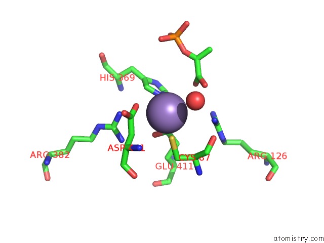
Mono view
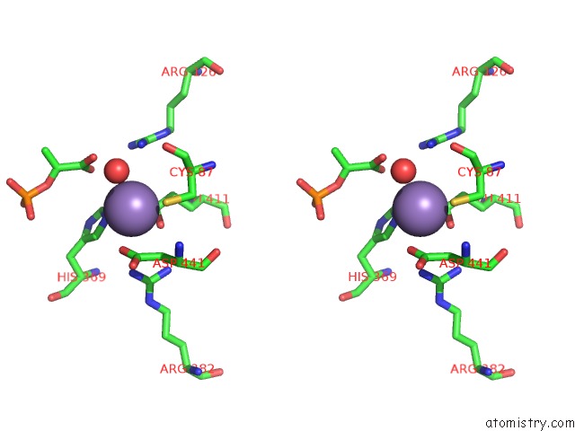
Stereo pair view
Mono view
Stereo pair view
A full contact list of Manganese with other atoms in the Mn binding
site number 2 of The Structure of 3-Deoxy-D-Arabino-Heptulosonate 7-Phosphate Synthase From Mycobacterium Tuberculosis within 5.0Å range:
|
Reference:
C.J.Webby,
H.M.Baker,
J.S.Lott,
E.N.Baker,
E.J.Parker.
The Structure of 3-Deoxy-D-Arabino-Heptulosonate 7-Phosphate Synthase From Mycobacterium Tuberculosis Reveals A Common Catalytic Scaffold and Ancestry For Type I and Type II Enzymes J.Mol.Biol. V. 354 927 2005.
ISSN: ISSN 0022-2836
PubMed: 16288916
DOI: 10.1016/J.JMB.2005.09.093
Page generated: Sat Oct 5 13:32:15 2024
ISSN: ISSN 0022-2836
PubMed: 16288916
DOI: 10.1016/J.JMB.2005.09.093
Last articles
Zn in 9MJ5Zn in 9HNW
Zn in 9G0L
Zn in 9FNE
Zn in 9DZN
Zn in 9E0I
Zn in 9D32
Zn in 9DAK
Zn in 8ZXC
Zn in 8ZUF