Manganese »
PDB 1yw7-2a2t »
1zql »
Manganese in PDB 1zql: Dna Polymerase Beta (Pol B) (E.C.2.7.7.7) Complexed with Seven Base Pairs of Dna; Soaked in the Presence of MNCL2 (15 Millimolar) and MGCL2 (15 Millimolar)
Protein crystallography data
The structure of Dna Polymerase Beta (Pol B) (E.C.2.7.7.7) Complexed with Seven Base Pairs of Dna; Soaked in the Presence of MNCL2 (15 Millimolar) and MGCL2 (15 Millimolar), PDB code: 1zql
was solved by
H.Pelletier,
M.R.Sawaya,
with X-Ray Crystallography technique. A brief refinement statistics is given in the table below:
| Resolution Low / High (Å) | 20.00 / 3.30 |
| Space group | P 21 21 2 |
| Cell size a, b, c (Å), α, β, γ (°) | 178.509, 57.730, 48.028, 90.00, 90.00, 90.00 |
| R / Rfree (%) | 15.9 / n/a |
Manganese Binding Sites:
The binding sites of Manganese atom in the Dna Polymerase Beta (Pol B) (E.C.2.7.7.7) Complexed with Seven Base Pairs of Dna; Soaked in the Presence of MNCL2 (15 Millimolar) and MGCL2 (15 Millimolar)
(pdb code 1zql). This binding sites where shown within
5.0 Angstroms radius around Manganese atom.
In total 3 binding sites of Manganese where determined in the Dna Polymerase Beta (Pol B) (E.C.2.7.7.7) Complexed with Seven Base Pairs of Dna; Soaked in the Presence of MNCL2 (15 Millimolar) and MGCL2 (15 Millimolar), PDB code: 1zql:
Jump to Manganese binding site number: 1; 2; 3;
In total 3 binding sites of Manganese where determined in the Dna Polymerase Beta (Pol B) (E.C.2.7.7.7) Complexed with Seven Base Pairs of Dna; Soaked in the Presence of MNCL2 (15 Millimolar) and MGCL2 (15 Millimolar), PDB code: 1zql:
Jump to Manganese binding site number: 1; 2; 3;
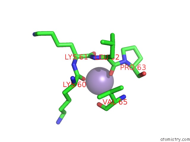
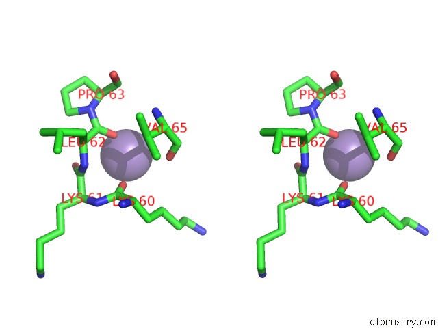
Manganese binding site 1 out of 3 in 1zql
Go back to
Manganese binding site 1 out
of 3 in the Dna Polymerase Beta (Pol B) (E.C.2.7.7.7) Complexed with Seven Base Pairs of Dna; Soaked in the Presence of MNCL2 (15 Millimolar) and MGCL2 (15 Millimolar)
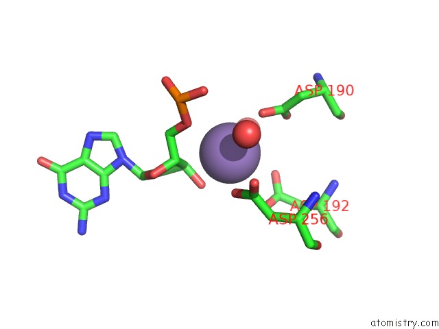
Mono view
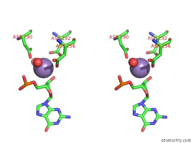
Stereo pair view
Mono view
Stereo pair view
A full contact list of Manganese with other atoms in the Mn binding
site number 1 of Dna Polymerase Beta (Pol B) (E.C.2.7.7.7) Complexed with Seven Base Pairs of Dna; Soaked in the Presence of MNCL2 (15 Millimolar) and MGCL2 (15 Millimolar) within 5.0Å range:
|
Manganese binding site 2 out of 3 in 1zql
Go back to
Manganese binding site 2 out
of 3 in the Dna Polymerase Beta (Pol B) (E.C.2.7.7.7) Complexed with Seven Base Pairs of Dna; Soaked in the Presence of MNCL2 (15 Millimolar) and MGCL2 (15 Millimolar)
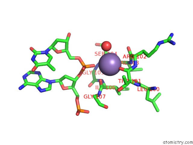
Mono view
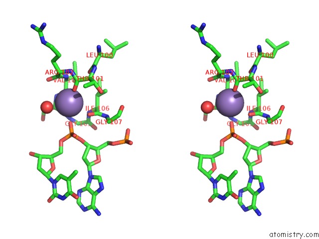
Stereo pair view
Mono view
Stereo pair view
A full contact list of Manganese with other atoms in the Mn binding
site number 2 of Dna Polymerase Beta (Pol B) (E.C.2.7.7.7) Complexed with Seven Base Pairs of Dna; Soaked in the Presence of MNCL2 (15 Millimolar) and MGCL2 (15 Millimolar) within 5.0Å range:
|
Manganese binding site 3 out of 3 in 1zql
Go back to
Manganese binding site 3 out
of 3 in the Dna Polymerase Beta (Pol B) (E.C.2.7.7.7) Complexed with Seven Base Pairs of Dna; Soaked in the Presence of MNCL2 (15 Millimolar) and MGCL2 (15 Millimolar)
Mono view
Stereo pair view
Mono view
Stereo pair view
A full contact list of Manganese with other atoms in the Mn binding
site number 3 of Dna Polymerase Beta (Pol B) (E.C.2.7.7.7) Complexed with Seven Base Pairs of Dna; Soaked in the Presence of MNCL2 (15 Millimolar) and MGCL2 (15 Millimolar) within 5.0Å range:
|
Reference:
H.Pelletier,
M.R.Sawaya.
Characterization of the Metal Ion Binding Helix-Hairpin-Helix Motifs in Human Dna Polymerase Beta By X-Ray Structural Analysis. Biochemistry V. 35 12778 1996.
ISSN: ISSN 0006-2960
PubMed: 8841120
DOI: 10.1021/BI960790I
Page generated: Sat Oct 5 13:18:15 2024
ISSN: ISSN 0006-2960
PubMed: 8841120
DOI: 10.1021/BI960790I
Last articles
Fe in 2YXOFe in 2YRS
Fe in 2YXC
Fe in 2YNM
Fe in 2YVJ
Fe in 2YP1
Fe in 2YU2
Fe in 2YU1
Fe in 2YQB
Fe in 2YOO