Manganese »
PDB 1yw7-2a2t »
1zj2 »
Manganese in PDB 1zj2: Crystal Structure of Human Galactosyltransferase (Gtb) Complexed with H Type I Trisaccharide
Protein crystallography data
The structure of Crystal Structure of Human Galactosyltransferase (Gtb) Complexed with H Type I Trisaccharide, PDB code: 1zj2
was solved by
J.A.Letts,
N.L.Rose,
Y.R.Fang,
C.H.Barry,
S.N.Borisova,
N.O.Seto,
M.M.Palcic,
S.V.Evans,
with X-Ray Crystallography technique. A brief refinement statistics is given in the table below:
| Resolution Low / High (Å) | 20.00 / 1.69 |
| Space group | C 2 2 21 |
| Cell size a, b, c (Å), α, β, γ (°) | 52.600, 150.430, 79.610, 90.00, 90.00, 90.00 |
| R / Rfree (%) | 20.6 / 23.2 |
Other elements in 1zj2:
The structure of Crystal Structure of Human Galactosyltransferase (Gtb) Complexed with H Type I Trisaccharide also contains other interesting chemical elements:
| Mercury | (Hg) | 6 atoms |
| Chlorine | (Cl) | 1 atom |
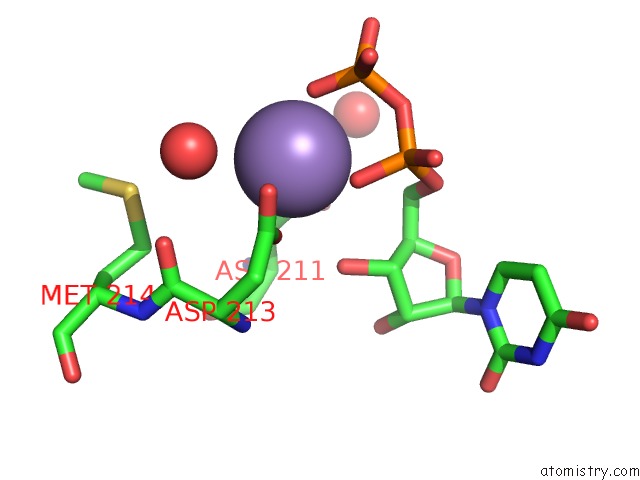
Manganese Binding Sites:
The binding sites of Manganese atom in the Crystal Structure of Human Galactosyltransferase (Gtb) Complexed with H Type I Trisaccharide
(pdb code 1zj2). This binding sites where shown within
5.0 Angstroms radius around Manganese atom.
In total only one binding site of Manganese was determined in the Crystal Structure of Human Galactosyltransferase (Gtb) Complexed with H Type I Trisaccharide, PDB code: 1zj2:
In total only one binding site of Manganese was determined in the Crystal Structure of Human Galactosyltransferase (Gtb) Complexed with H Type I Trisaccharide, PDB code: 1zj2:
Manganese binding site 1 out of 1 in 1zj2
Go back to
Manganese binding site 1 out
of 1 in the Crystal Structure of Human Galactosyltransferase (Gtb) Complexed with H Type I Trisaccharide
Mono view

Stereo pair view
Mono view
Stereo pair view
A full contact list of Manganese with other atoms in the Mn binding
site number 1 of Crystal Structure of Human Galactosyltransferase (Gtb) Complexed with H Type I Trisaccharide within 5.0Å range:
|
Reference:
J.A.Letts,
N.L.Rose,
Y.R.Fang,
C.H.Barry,
S.N.Borisova,
N.O.Seto,
M.M.Palcic,
S.V.Evans.
Differential Recognition of the Type I and II H Antigen Acceptors By the Human Abo(H) Blood Group A and B Glycosyltransferases. J.Biol.Chem. V. 281 3625 2006.
ISSN: ISSN 0021-9258
PubMed: 16326711
DOI: 10.1074/JBC.M507620200
Page generated: Sat Oct 5 13:17:36 2024
ISSN: ISSN 0021-9258
PubMed: 16326711
DOI: 10.1074/JBC.M507620200
Last articles
Ca in 5VCOCa in 5VCN
Ca in 5VC1
Ca in 5VA9
Ca in 5VBJ
Ca in 5VAO
Ca in 5V8K
Ca in 5V6V
Ca in 5V2C
Ca in 5V4D