Manganese »
PDB 1yw7-2a2t »
1za0 »
Manganese in PDB 1za0: X-Ray Structure of Putative Acyl-Acp Desaturase DESA2 From Mycobacterium Tuberculosis H37RV
Enzymatic activity of X-Ray Structure of Putative Acyl-Acp Desaturase DESA2 From Mycobacterium Tuberculosis H37RV
All present enzymatic activity of X-Ray Structure of Putative Acyl-Acp Desaturase DESA2 From Mycobacterium Tuberculosis H37RV:
1.14.19.2;
1.14.19.2;
Protein crystallography data
The structure of X-Ray Structure of Putative Acyl-Acp Desaturase DESA2 From Mycobacterium Tuberculosis H37RV, PDB code: 1za0
was solved by
H.D.Dyer,
K.S.Lyle,
I.Rayment,
B.G.Fox,
with X-Ray Crystallography technique. A brief refinement statistics is given in the table below:
| Resolution Low / High (Å) | 49.30 / 2.00 |
| Space group | P 65 2 2 |
| Cell size a, b, c (Å), α, β, γ (°) | 66.100, 66.100, 292.000, 90.00, 90.00, 120.00 |
| R / Rfree (%) | 20.6 / 23.7 |
Manganese Binding Sites:
The binding sites of Manganese atom in the X-Ray Structure of Putative Acyl-Acp Desaturase DESA2 From Mycobacterium Tuberculosis H37RV
(pdb code 1za0). This binding sites where shown within
5.0 Angstroms radius around Manganese atom.
In total only one binding site of Manganese was determined in the X-Ray Structure of Putative Acyl-Acp Desaturase DESA2 From Mycobacterium Tuberculosis H37RV, PDB code: 1za0:
In total only one binding site of Manganese was determined in the X-Ray Structure of Putative Acyl-Acp Desaturase DESA2 From Mycobacterium Tuberculosis H37RV, PDB code: 1za0:
Manganese binding site 1 out of 1 in 1za0
Go back to
Manganese binding site 1 out
of 1 in the X-Ray Structure of Putative Acyl-Acp Desaturase DESA2 From Mycobacterium Tuberculosis H37RV
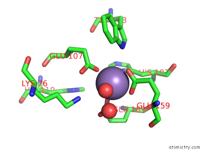
Mono view
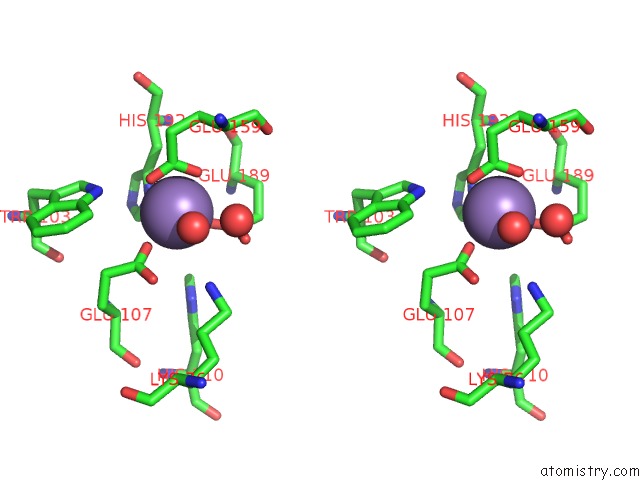
Stereo pair view
Mono view
Stereo pair view
A full contact list of Manganese with other atoms in the Mn binding
site number 1 of X-Ray Structure of Putative Acyl-Acp Desaturase DESA2 From Mycobacterium Tuberculosis H37RV within 5.0Å range:
|
Reference:
D.H.Dyer,
K.S.Lyle,
I.Rayment,
B.G.Fox.
X-Ray Structure of Putative Acyl-Acp Desaturase DESA2 From Mycobacterium Tuberculosis H37RV. Protein Sci. V. 14 1508 2005.
ISSN: ISSN 0961-8368
PubMed: 15929999
DOI: 10.1110/PS.041288005
Page generated: Sat Oct 5 13:16:20 2024
ISSN: ISSN 0961-8368
PubMed: 15929999
DOI: 10.1110/PS.041288005
Last articles
Zn in 9J0NZn in 9J0O
Zn in 9J0P
Zn in 9FJX
Zn in 9EKB
Zn in 9C0F
Zn in 9CAH
Zn in 9CH0
Zn in 9CH3
Zn in 9CH1