Manganese »
PDB 1xif-1ytm »
1yny »
Manganese in PDB 1yny: Molecular Structure of D-Hydantoinase From A Bacillus Sp. AR9: Evidence For Mercury Inhibition
Enzymatic activity of Molecular Structure of D-Hydantoinase From A Bacillus Sp. AR9: Evidence For Mercury Inhibition
All present enzymatic activity of Molecular Structure of D-Hydantoinase From A Bacillus Sp. AR9: Evidence For Mercury Inhibition:
3.5.2.2;
3.5.2.2;
Protein crystallography data
The structure of Molecular Structure of D-Hydantoinase From A Bacillus Sp. AR9: Evidence For Mercury Inhibition, PDB code: 1yny
was solved by
K.V.Radha Kishan,
R.M.Vohra,
K.Ganeshan,
V.Agrawal,
V.M.Sharma,
R.Sharma,
with X-Ray Crystallography technique. A brief refinement statistics is given in the table below:
| Resolution Low / High (Å) | 50.00 / 2.30 |
| Space group | P 64 |
| Cell size a, b, c (Å), α, β, γ (°) | 129.540, 129.540, 102.850, 90.00, 90.00, 120.00 |
| R / Rfree (%) | 19.9 / 24.3 |
Manganese Binding Sites:
The binding sites of Manganese atom in the Molecular Structure of D-Hydantoinase From A Bacillus Sp. AR9: Evidence For Mercury Inhibition
(pdb code 1yny). This binding sites where shown within
5.0 Angstroms radius around Manganese atom.
In total 4 binding sites of Manganese where determined in the Molecular Structure of D-Hydantoinase From A Bacillus Sp. AR9: Evidence For Mercury Inhibition, PDB code: 1yny:
Jump to Manganese binding site number: 1; 2; 3; 4;
In total 4 binding sites of Manganese where determined in the Molecular Structure of D-Hydantoinase From A Bacillus Sp. AR9: Evidence For Mercury Inhibition, PDB code: 1yny:
Jump to Manganese binding site number: 1; 2; 3; 4;
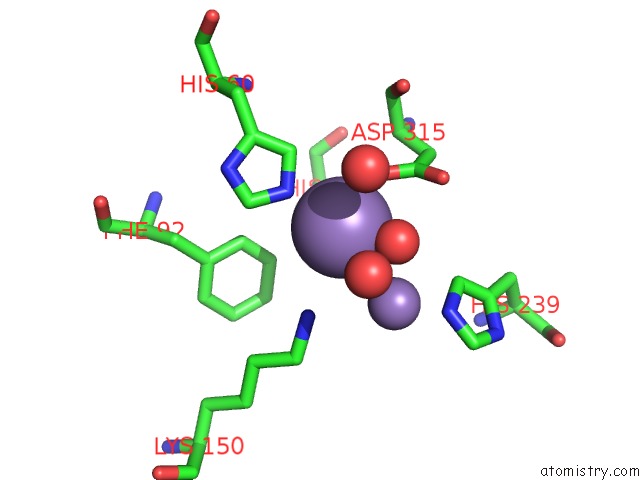
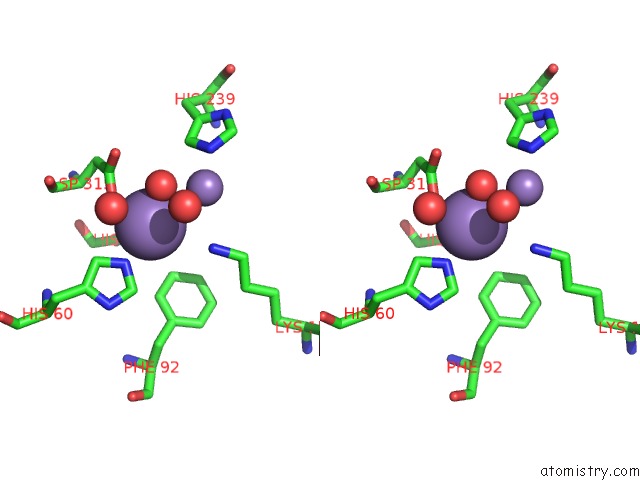
Manganese binding site 1 out of 4 in 1yny
Go back to
Manganese binding site 1 out
of 4 in the Molecular Structure of D-Hydantoinase From A Bacillus Sp. AR9: Evidence For Mercury Inhibition
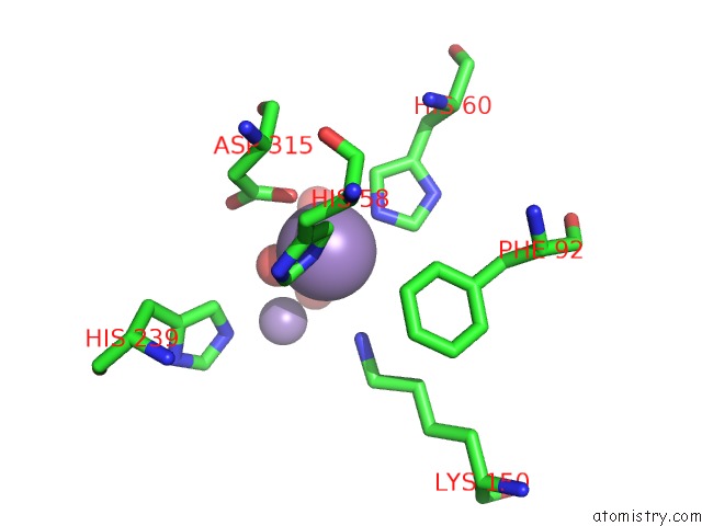
Mono view
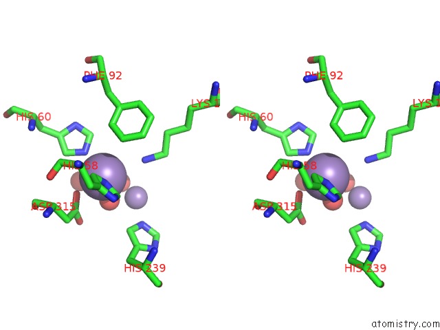
Stereo pair view
Mono view
Stereo pair view
A full contact list of Manganese with other atoms in the Mn binding
site number 1 of Molecular Structure of D-Hydantoinase From A Bacillus Sp. AR9: Evidence For Mercury Inhibition within 5.0Å range:
|
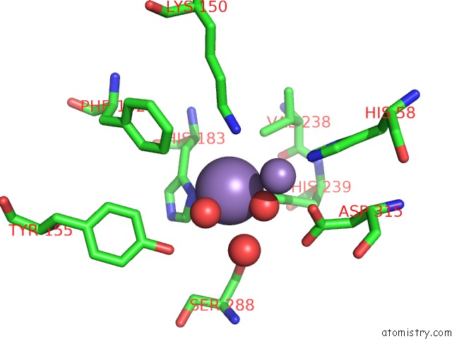
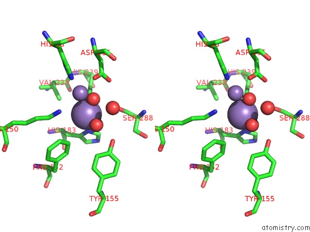
Manganese binding site 2 out of 4 in 1yny
Go back to
Manganese binding site 2 out
of 4 in the Molecular Structure of D-Hydantoinase From A Bacillus Sp. AR9: Evidence For Mercury Inhibition
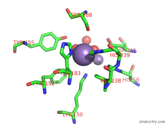
Mono view
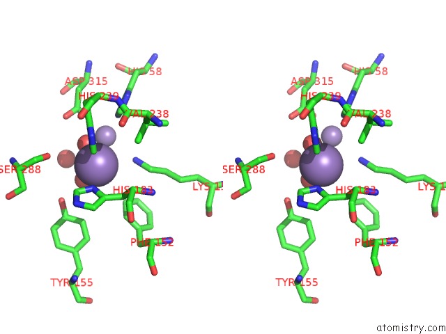
Stereo pair view
Mono view
Stereo pair view
A full contact list of Manganese with other atoms in the Mn binding
site number 2 of Molecular Structure of D-Hydantoinase From A Bacillus Sp. AR9: Evidence For Mercury Inhibition within 5.0Å range:
|
Manganese binding site 3 out of 4 in 1yny
Go back to
Manganese binding site 3 out
of 4 in the Molecular Structure of D-Hydantoinase From A Bacillus Sp. AR9: Evidence For Mercury Inhibition
Mono view
Stereo pair view
Mono view
Stereo pair view
A full contact list of Manganese with other atoms in the Mn binding
site number 3 of Molecular Structure of D-Hydantoinase From A Bacillus Sp. AR9: Evidence For Mercury Inhibition within 5.0Å range:
|
Manganese binding site 4 out of 4 in 1yny
Go back to
Manganese binding site 4 out
of 4 in the Molecular Structure of D-Hydantoinase From A Bacillus Sp. AR9: Evidence For Mercury Inhibition
Mono view
Stereo pair view
Mono view
Stereo pair view
A full contact list of Manganese with other atoms in the Mn binding
site number 4 of Molecular Structure of D-Hydantoinase From A Bacillus Sp. AR9: Evidence For Mercury Inhibition within 5.0Å range:
|
Reference:
K.V.Radha Kishan,
R.M.Vohra,
K.Ganesan,
V.Agrawal,
V.M.Sharma,
R.Sharma.
Molecular Structure of D-Hydantoinase From Bacillus Sp. AR9: Evidence For Mercury Inhibition. J.Mol.Biol. V. 347 95 2005.
ISSN: ISSN 0022-2836
PubMed: 15733920
DOI: 10.1016/J.JMB.2005.01.025
Page generated: Sat Oct 5 13:13:17 2024
ISSN: ISSN 0022-2836
PubMed: 15733920
DOI: 10.1016/J.JMB.2005.01.025
Last articles
Zn in 9J0NZn in 9J0O
Zn in 9J0P
Zn in 9FJX
Zn in 9EKB
Zn in 9C0F
Zn in 9CAH
Zn in 9CH0
Zn in 9CH3
Zn in 9CH1