Manganese »
PDB 1xif-1ytm »
1ylh »
Manganese in PDB 1ylh: Crystal Structure of Phosphoenolpyruvate Carboxykinase From Actinobaccilus Succinogenes in Complex with Manganese and Pyruvate
Enzymatic activity of Crystal Structure of Phosphoenolpyruvate Carboxykinase From Actinobaccilus Succinogenes in Complex with Manganese and Pyruvate
All present enzymatic activity of Crystal Structure of Phosphoenolpyruvate Carboxykinase From Actinobaccilus Succinogenes in Complex with Manganese and Pyruvate:
4.1.1.49;
4.1.1.49;
Protein crystallography data
The structure of Crystal Structure of Phosphoenolpyruvate Carboxykinase From Actinobaccilus Succinogenes in Complex with Manganese and Pyruvate, PDB code: 1ylh
was solved by
Y.A.Leduc,
L.Prasad,
M.Laivenieks,
J.G.Zeikus,
L.T.Delbaere,
with X-Ray Crystallography technique. A brief refinement statistics is given in the table below:
| Resolution Low / High (Å) | 26.26 / 1.70 |
| Space group | P 1 21 1 |
| Cell size a, b, c (Å), α, β, γ (°) | 56.737, 55.089, 90.077, 90.00, 106.17, 90.00 |
| R / Rfree (%) | 19 / 21.5 |
Manganese Binding Sites:
The binding sites of Manganese atom in the Crystal Structure of Phosphoenolpyruvate Carboxykinase From Actinobaccilus Succinogenes in Complex with Manganese and Pyruvate
(pdb code 1ylh). This binding sites where shown within
5.0 Angstroms radius around Manganese atom.
In total only one binding site of Manganese was determined in the Crystal Structure of Phosphoenolpyruvate Carboxykinase From Actinobaccilus Succinogenes in Complex with Manganese and Pyruvate, PDB code: 1ylh:
In total only one binding site of Manganese was determined in the Crystal Structure of Phosphoenolpyruvate Carboxykinase From Actinobaccilus Succinogenes in Complex with Manganese and Pyruvate, PDB code: 1ylh:
Manganese binding site 1 out of 1 in 1ylh
Go back to
Manganese binding site 1 out
of 1 in the Crystal Structure of Phosphoenolpyruvate Carboxykinase From Actinobaccilus Succinogenes in Complex with Manganese and Pyruvate
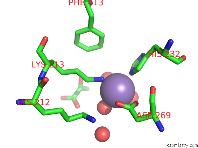
Mono view
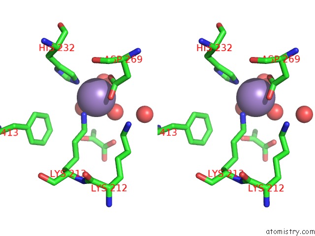
Stereo pair view
Mono view
Stereo pair view
A full contact list of Manganese with other atoms in the Mn binding
site number 1 of Crystal Structure of Phosphoenolpyruvate Carboxykinase From Actinobaccilus Succinogenes in Complex with Manganese and Pyruvate within 5.0Å range:
|
Reference:
Y.A.Leduc,
L.Prasad,
M.Laivenieks,
J.G.Zeikus,
L.T.Delbaere.
Structure of Pep Carboxykinase From the Succinate-Producing Actinobacillus Succinogenes: A New Conserved Active-Site Motif. Acta Crystallogr.,Sect.D V. 61 903 2005.
ISSN: ISSN 0907-4449
PubMed: 15983413
DOI: 10.1107/S0907444905008723
Page generated: Sat Oct 5 13:12:33 2024
ISSN: ISSN 0907-4449
PubMed: 15983413
DOI: 10.1107/S0907444905008723
Last articles
Zn in 9J0NZn in 9J0O
Zn in 9J0P
Zn in 9FJX
Zn in 9EKB
Zn in 9C0F
Zn in 9CAH
Zn in 9CH0
Zn in 9CH3
Zn in 9CH1