Manganese »
PDB 1xif-1ytm »
1ybu »
Manganese in PDB 1ybu: Mycobacterium Tuberculosis Adenylyl Cyclase RV1900C Chd, in Complex with A Substrate Analog.
Enzymatic activity of Mycobacterium Tuberculosis Adenylyl Cyclase RV1900C Chd, in Complex with A Substrate Analog.
All present enzymatic activity of Mycobacterium Tuberculosis Adenylyl Cyclase RV1900C Chd, in Complex with A Substrate Analog.:
4.6.1.1;
4.6.1.1;
Protein crystallography data
The structure of Mycobacterium Tuberculosis Adenylyl Cyclase RV1900C Chd, in Complex with A Substrate Analog., PDB code: 1ybu
was solved by
S.C.Sinha,
M.Wetterer,
S.R.Sprang,
J.E.Schultz,
J.U.Linder,
with X-Ray Crystallography technique. A brief refinement statistics is given in the table below:
| Resolution Low / High (Å) | 31.94 / 2.40 |
| Space group | P 1 2 1 |
| Cell size a, b, c (Å), α, β, γ (°) | 91.067, 48.923, 68.198, 90.00, 90.00, 90.00 |
| R / Rfree (%) | 23.3 / 26.2 |
Manganese Binding Sites:
The binding sites of Manganese atom in the Mycobacterium Tuberculosis Adenylyl Cyclase RV1900C Chd, in Complex with A Substrate Analog.
(pdb code 1ybu). This binding sites where shown within
5.0 Angstroms radius around Manganese atom.
In total 2 binding sites of Manganese where determined in the Mycobacterium Tuberculosis Adenylyl Cyclase RV1900C Chd, in Complex with A Substrate Analog., PDB code: 1ybu:
Jump to Manganese binding site number: 1; 2;
In total 2 binding sites of Manganese where determined in the Mycobacterium Tuberculosis Adenylyl Cyclase RV1900C Chd, in Complex with A Substrate Analog., PDB code: 1ybu:
Jump to Manganese binding site number: 1; 2;
Manganese binding site 1 out of 2 in 1ybu
Go back to
Manganese binding site 1 out
of 2 in the Mycobacterium Tuberculosis Adenylyl Cyclase RV1900C Chd, in Complex with A Substrate Analog.
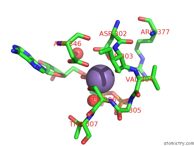
Mono view
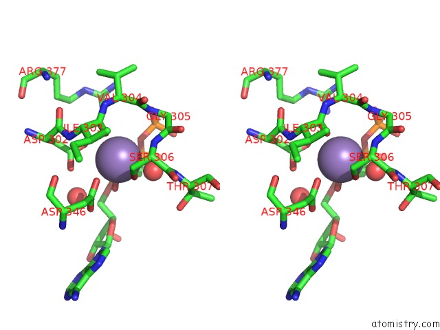
Stereo pair view
Mono view
Stereo pair view
A full contact list of Manganese with other atoms in the Mn binding
site number 1 of Mycobacterium Tuberculosis Adenylyl Cyclase RV1900C Chd, in Complex with A Substrate Analog. within 5.0Å range:
|
Manganese binding site 2 out of 2 in 1ybu
Go back to
Manganese binding site 2 out
of 2 in the Mycobacterium Tuberculosis Adenylyl Cyclase RV1900C Chd, in Complex with A Substrate Analog.
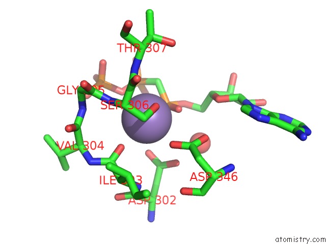
Mono view
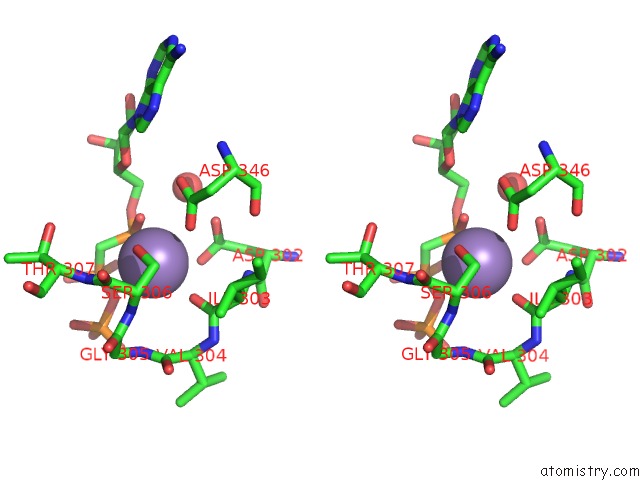
Stereo pair view
Mono view
Stereo pair view
A full contact list of Manganese with other atoms in the Mn binding
site number 2 of Mycobacterium Tuberculosis Adenylyl Cyclase RV1900C Chd, in Complex with A Substrate Analog. within 5.0Å range:
|
Reference:
S.C.Sinha,
M.Wetterer,
S.R.Sprang,
J.E.Schultz,
J.U.Linder.
Origin of Asymmetry in Adenylyl Cyclases: Structures of Mycobacterium Tuberculosis RV1900C. Embo J. V. 24 663 2005.
ISSN: ISSN 0261-4189
PubMed: 15678099
DOI: 10.1038/SJ.EMBOJ.7600573
Page generated: Sat Oct 5 13:10:26 2024
ISSN: ISSN 0261-4189
PubMed: 15678099
DOI: 10.1038/SJ.EMBOJ.7600573
Last articles
Zn in 9J0NZn in 9J0O
Zn in 9J0P
Zn in 9FJX
Zn in 9EKB
Zn in 9C0F
Zn in 9CAH
Zn in 9CH0
Zn in 9CH3
Zn in 9CH1