Manganese »
PDB 1xif-1ytm »
1xmf »
Manganese in PDB 1xmf: Structure of Mn(II)-Soaked Apo Methane Monooxygenase Hydroxylase Crystals From M. Capsulatus (Bath)
Enzymatic activity of Structure of Mn(II)-Soaked Apo Methane Monooxygenase Hydroxylase Crystals From M. Capsulatus (Bath)
All present enzymatic activity of Structure of Mn(II)-Soaked Apo Methane Monooxygenase Hydroxylase Crystals From M. Capsulatus (Bath):
1.14.13.25;
1.14.13.25;
Protein crystallography data
The structure of Structure of Mn(II)-Soaked Apo Methane Monooxygenase Hydroxylase Crystals From M. Capsulatus (Bath), PDB code: 1xmf
was solved by
M.H.Sazinsky,
M.Merkx,
E.Cadieux,
S.Tang,
S.J.Lippard,
with X-Ray Crystallography technique. A brief refinement statistics is given in the table below:
| Resolution Low / High (Å) | 28.76 / 2.32 |
| Space group | P 21 21 21 |
| Cell size a, b, c (Å), α, β, γ (°) | 70.820, 171.679, 220.275, 90.00, 90.00, 90.00 |
| R / Rfree (%) | 21.9 / 26.3 |
Other elements in 1xmf:
The structure of Structure of Mn(II)-Soaked Apo Methane Monooxygenase Hydroxylase Crystals From M. Capsulatus (Bath) also contains other interesting chemical elements:
| Calcium | (Ca) | 4 atoms |
Manganese Binding Sites:
The binding sites of Manganese atom in the Structure of Mn(II)-Soaked Apo Methane Monooxygenase Hydroxylase Crystals From M. Capsulatus (Bath)
(pdb code 1xmf). This binding sites where shown within
5.0 Angstroms radius around Manganese atom.
In total 4 binding sites of Manganese where determined in the Structure of Mn(II)-Soaked Apo Methane Monooxygenase Hydroxylase Crystals From M. Capsulatus (Bath), PDB code: 1xmf:
Jump to Manganese binding site number: 1; 2; 3; 4;
In total 4 binding sites of Manganese where determined in the Structure of Mn(II)-Soaked Apo Methane Monooxygenase Hydroxylase Crystals From M. Capsulatus (Bath), PDB code: 1xmf:
Jump to Manganese binding site number: 1; 2; 3; 4;
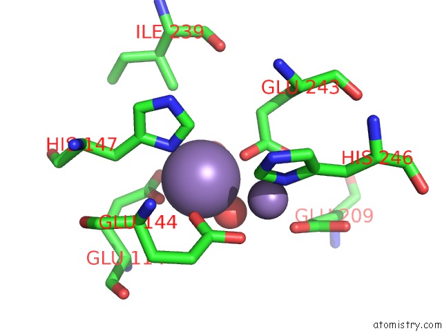
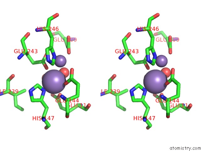
Manganese binding site 1 out of 4 in 1xmf
Go back to
Manganese binding site 1 out
of 4 in the Structure of Mn(II)-Soaked Apo Methane Monooxygenase Hydroxylase Crystals From M. Capsulatus (Bath)
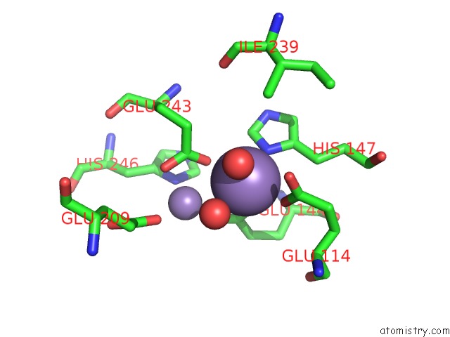
Mono view
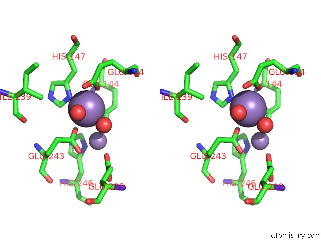
Stereo pair view
Mono view
Stereo pair view
A full contact list of Manganese with other atoms in the Mn binding
site number 1 of Structure of Mn(II)-Soaked Apo Methane Monooxygenase Hydroxylase Crystals From M. Capsulatus (Bath) within 5.0Å range:
|
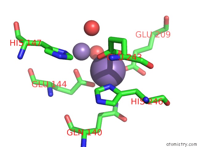
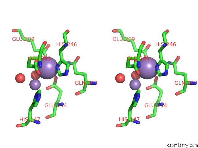
Manganese binding site 2 out of 4 in 1xmf
Go back to
Manganese binding site 2 out
of 4 in the Structure of Mn(II)-Soaked Apo Methane Monooxygenase Hydroxylase Crystals From M. Capsulatus (Bath)
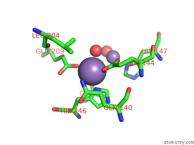
Mono view
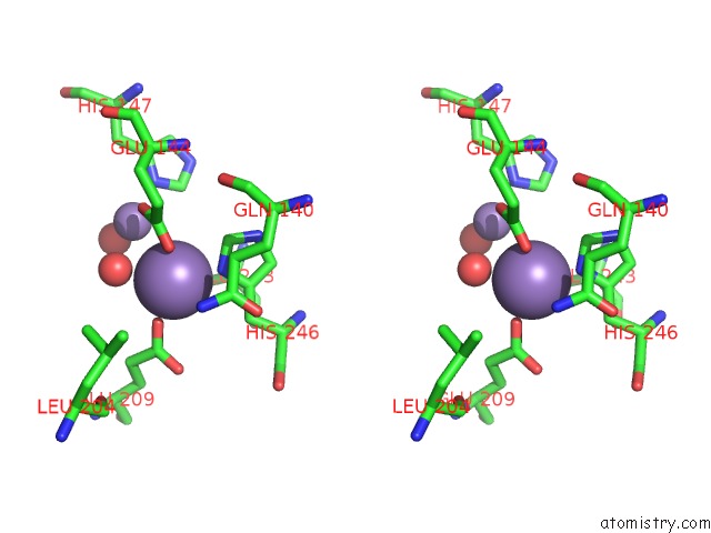
Stereo pair view
Mono view
Stereo pair view
A full contact list of Manganese with other atoms in the Mn binding
site number 2 of Structure of Mn(II)-Soaked Apo Methane Monooxygenase Hydroxylase Crystals From M. Capsulatus (Bath) within 5.0Å range:
|
Manganese binding site 3 out of 4 in 1xmf
Go back to
Manganese binding site 3 out
of 4 in the Structure of Mn(II)-Soaked Apo Methane Monooxygenase Hydroxylase Crystals From M. Capsulatus (Bath)
Mono view
Stereo pair view
Mono view
Stereo pair view
A full contact list of Manganese with other atoms in the Mn binding
site number 3 of Structure of Mn(II)-Soaked Apo Methane Monooxygenase Hydroxylase Crystals From M. Capsulatus (Bath) within 5.0Å range:
|
Manganese binding site 4 out of 4 in 1xmf
Go back to
Manganese binding site 4 out
of 4 in the Structure of Mn(II)-Soaked Apo Methane Monooxygenase Hydroxylase Crystals From M. Capsulatus (Bath)
Mono view
Stereo pair view
Mono view
Stereo pair view
A full contact list of Manganese with other atoms in the Mn binding
site number 4 of Structure of Mn(II)-Soaked Apo Methane Monooxygenase Hydroxylase Crystals From M. Capsulatus (Bath) within 5.0Å range:
|
Reference:
M.H.Sazinsky,
M.Merkx,
E.Cadieux,
S.Tang,
S.J.Lippard.
Preparation and X-Ray Structures of Metal-Free, Dicobalt and Dimanganese Forms of Soluble Methane Monooxygenase Hydroxylase From Methylococcus Capsulatus (Bath) Biochemistry V. 43 16263 2004.
ISSN: ISSN 0006-2960
PubMed: 15610020
DOI: 10.1021/BI048140Z
Page generated: Sat Oct 5 13:07:29 2024
ISSN: ISSN 0006-2960
PubMed: 15610020
DOI: 10.1021/BI048140Z
Last articles
Ca in 5NGQCa in 5NH5
Ca in 5NGY
Ca in 5NG1
Ca in 5NFZ
Ca in 5NF3
Ca in 5NF2
Ca in 5NF0
Ca in 5NEY
Ca in 5NES