Manganese »
PDB 1w2c-1xie »
1wsf »
Manganese in PDB 1wsf: Co-Crystal Structure of E.Coli Rnase Hi Active Site Mutant (D134A*) with MN2+
Enzymatic activity of Co-Crystal Structure of E.Coli Rnase Hi Active Site Mutant (D134A*) with MN2+
All present enzymatic activity of Co-Crystal Structure of E.Coli Rnase Hi Active Site Mutant (D134A*) with MN2+:
3.1.26.4;
3.1.26.4;
Protein crystallography data
The structure of Co-Crystal Structure of E.Coli Rnase Hi Active Site Mutant (D134A*) with MN2+, PDB code: 1wsf
was solved by
Y.Tsunaka,
K.Takano,
H.Matsumura,
Y.Yamagata,
S.Kanaya,
with X-Ray Crystallography technique. A brief refinement statistics is given in the table below:
| Resolution Low / High (Å) | 50.00 / 2.30 |
| Space group | P 21 21 2 |
| Cell size a, b, c (Å), α, β, γ (°) | 120.990, 80.089, 63.951, 90.00, 90.00, 90.00 |
| R / Rfree (%) | 21.6 / 29 |
Manganese Binding Sites:
The binding sites of Manganese atom in the Co-Crystal Structure of E.Coli Rnase Hi Active Site Mutant (D134A*) with MN2+
(pdb code 1wsf). This binding sites where shown within
5.0 Angstroms radius around Manganese atom.
In total 3 binding sites of Manganese where determined in the Co-Crystal Structure of E.Coli Rnase Hi Active Site Mutant (D134A*) with MN2+, PDB code: 1wsf:
Jump to Manganese binding site number: 1; 2; 3;
In total 3 binding sites of Manganese where determined in the Co-Crystal Structure of E.Coli Rnase Hi Active Site Mutant (D134A*) with MN2+, PDB code: 1wsf:
Jump to Manganese binding site number: 1; 2; 3;
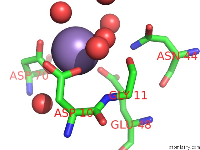
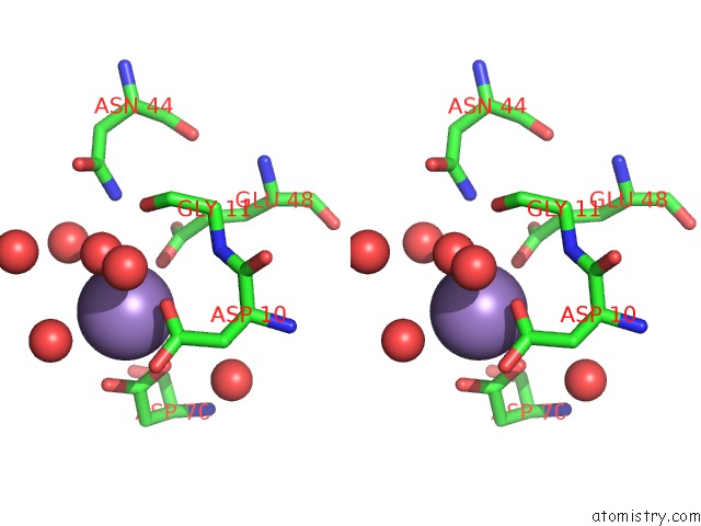
Manganese binding site 1 out of 3 in 1wsf
Go back to
Manganese binding site 1 out
of 3 in the Co-Crystal Structure of E.Coli Rnase Hi Active Site Mutant (D134A*) with MN2+
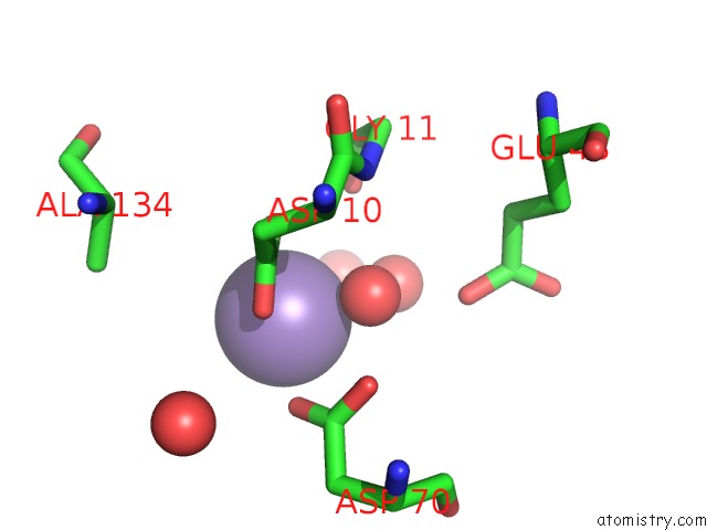
Mono view
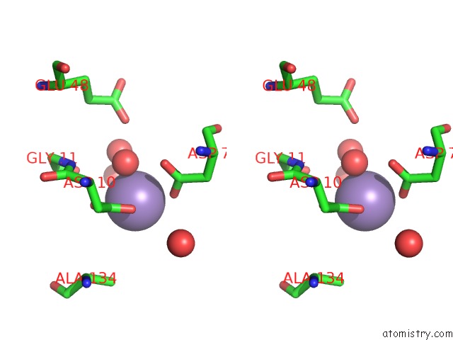
Stereo pair view
Mono view
Stereo pair view
A full contact list of Manganese with other atoms in the Mn binding
site number 1 of Co-Crystal Structure of E.Coli Rnase Hi Active Site Mutant (D134A*) with MN2+ within 5.0Å range:
|
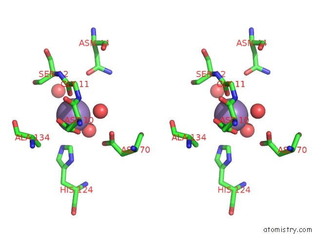
Manganese binding site 2 out of 3 in 1wsf
Go back to
Manganese binding site 2 out
of 3 in the Co-Crystal Structure of E.Coli Rnase Hi Active Site Mutant (D134A*) with MN2+
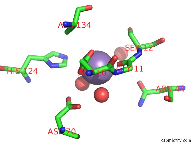
Mono view
Stereo pair view
Mono view
Stereo pair view
A full contact list of Manganese with other atoms in the Mn binding
site number 2 of Co-Crystal Structure of E.Coli Rnase Hi Active Site Mutant (D134A*) with MN2+ within 5.0Å range:
|
Manganese binding site 3 out of 3 in 1wsf
Go back to
Manganese binding site 3 out
of 3 in the Co-Crystal Structure of E.Coli Rnase Hi Active Site Mutant (D134A*) with MN2+
Mono view
Stereo pair view
Mono view
Stereo pair view
A full contact list of Manganese with other atoms in the Mn binding
site number 3 of Co-Crystal Structure of E.Coli Rnase Hi Active Site Mutant (D134A*) with MN2+ within 5.0Å range:
|
Reference:
Y.Tsunaka,
K.Takano,
H.Matsumura,
Y.Yamagata,
S.Kanaya.
Identification of Single Mn(2+) Binding Sites Required For Activation of the Mutant Proteins of E.Coli Rnase Hi at GLU48 and/or ASP134 By X-Ray Crystallography J.Mol.Biol. V. 345 1171 2005.
ISSN: ISSN 0022-2836
PubMed: 15644213
DOI: 10.1016/J.JMB.2004.11.007
Page generated: Sat Oct 5 12:58:29 2024
ISSN: ISSN 0022-2836
PubMed: 15644213
DOI: 10.1016/J.JMB.2004.11.007
Last articles
F in 7M03F in 7M02
F in 7LZU
F in 7LZW
F in 7LY8
F in 7LWG
F in 7LZV
F in 7LZF
F in 7LZD
F in 7LZA