Manganese »
PDB 1w2c-1xie »
1wse »
Manganese in PDB 1wse: Co-Crystal Structure of E.Coli Rnase Hi Active Site Mutant (E48A*) with MN2+
Enzymatic activity of Co-Crystal Structure of E.Coli Rnase Hi Active Site Mutant (E48A*) with MN2+
All present enzymatic activity of Co-Crystal Structure of E.Coli Rnase Hi Active Site Mutant (E48A*) with MN2+:
3.1.26.4;
3.1.26.4;
Protein crystallography data
The structure of Co-Crystal Structure of E.Coli Rnase Hi Active Site Mutant (E48A*) with MN2+, PDB code: 1wse
was solved by
Y.Tsunaka,
K.Takano,
H.Matsumura,
Y.Yamagata,
S.Kanaya,
with X-Ray Crystallography technique. A brief refinement statistics is given in the table below:
| Resolution Low / High (Å) | 50.00 / 2.30 |
| Space group | P 2 2 21 |
| Cell size a, b, c (Å), α, β, γ (°) | 58.041, 66.008, 79.485, 90.00, 90.00, 90.00 |
| R / Rfree (%) | 23 / 28.8 |
Manganese Binding Sites:
The binding sites of Manganese atom in the Co-Crystal Structure of E.Coli Rnase Hi Active Site Mutant (E48A*) with MN2+
(pdb code 1wse). This binding sites where shown within
5.0 Angstroms radius around Manganese atom.
In total 2 binding sites of Manganese where determined in the Co-Crystal Structure of E.Coli Rnase Hi Active Site Mutant (E48A*) with MN2+, PDB code: 1wse:
Jump to Manganese binding site number: 1; 2;
In total 2 binding sites of Manganese where determined in the Co-Crystal Structure of E.Coli Rnase Hi Active Site Mutant (E48A*) with MN2+, PDB code: 1wse:
Jump to Manganese binding site number: 1; 2;
Manganese binding site 1 out of 2 in 1wse
Go back to
Manganese binding site 1 out
of 2 in the Co-Crystal Structure of E.Coli Rnase Hi Active Site Mutant (E48A*) with MN2+
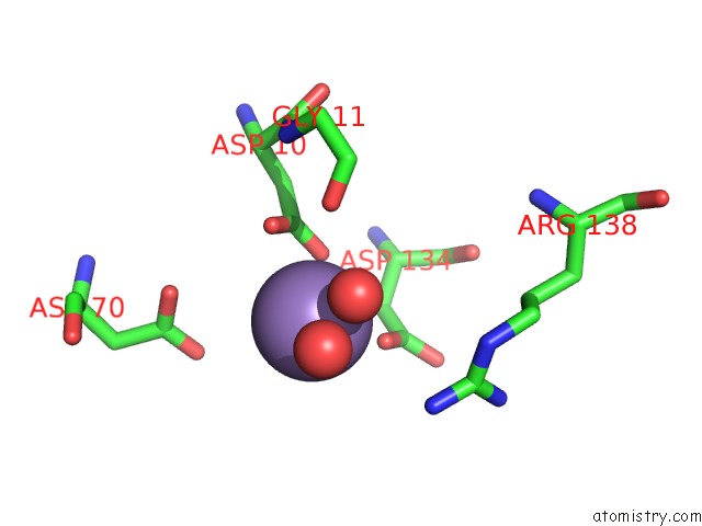
Mono view

Stereo pair view
Mono view
Stereo pair view
A full contact list of Manganese with other atoms in the Mn binding
site number 1 of Co-Crystal Structure of E.Coli Rnase Hi Active Site Mutant (E48A*) with MN2+ within 5.0Å range:
|
Manganese binding site 2 out of 2 in 1wse
Go back to
Manganese binding site 2 out
of 2 in the Co-Crystal Structure of E.Coli Rnase Hi Active Site Mutant (E48A*) with MN2+
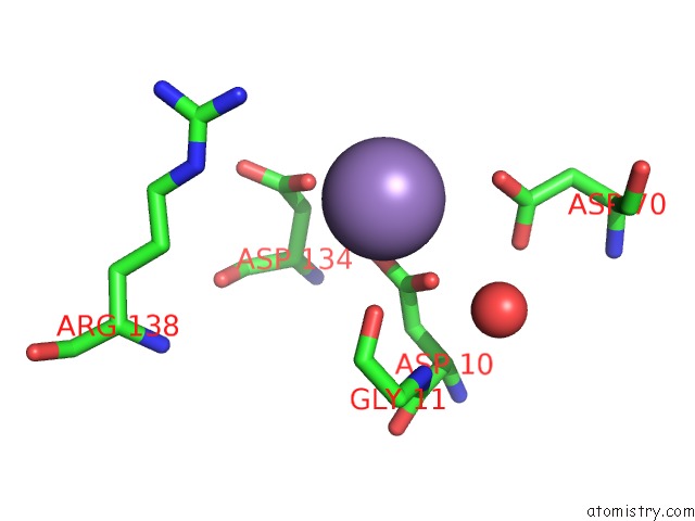
Mono view
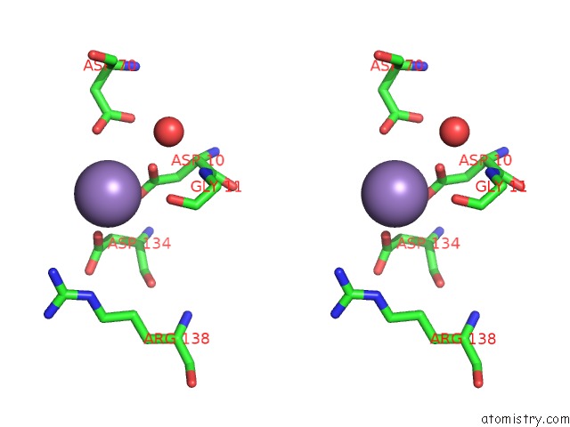
Stereo pair view
Mono view
Stereo pair view
A full contact list of Manganese with other atoms in the Mn binding
site number 2 of Co-Crystal Structure of E.Coli Rnase Hi Active Site Mutant (E48A*) with MN2+ within 5.0Å range:
|
Reference:
Y.Tsunaka,
K.Takano,
H.Matsumura,
Y.Yamagata,
S.Kanaya.
Identification of Single Mn(2+) Binding Sites Required For Activation of the Mutant Proteins of E.Coli Rnase Hi at GLU48 and/or ASP134 By X-Ray Crystallography J.Mol.Biol. V. 345 1171 2005.
ISSN: ISSN 0022-2836
PubMed: 15644213
DOI: 10.1016/J.JMB.2004.11.007
Page generated: Sat Oct 5 12:57:45 2024
ISSN: ISSN 0022-2836
PubMed: 15644213
DOI: 10.1016/J.JMB.2004.11.007
Last articles
Zn in 9MJ5Zn in 9HNW
Zn in 9G0L
Zn in 9FNE
Zn in 9DZN
Zn in 9E0I
Zn in 9D32
Zn in 9DAK
Zn in 8ZXC
Zn in 8ZUF