Manganese »
PDB 1w2c-1xie »
1woi »
Manganese in PDB 1woi: Crystal Structure of Agmatinase Reveals Structural Conservation and Inhibition Mechanism of the Ureohydrolase Superfamily
Enzymatic activity of Crystal Structure of Agmatinase Reveals Structural Conservation and Inhibition Mechanism of the Ureohydrolase Superfamily
All present enzymatic activity of Crystal Structure of Agmatinase Reveals Structural Conservation and Inhibition Mechanism of the Ureohydrolase Superfamily:
3.5.3.11;
3.5.3.11;
Protein crystallography data
The structure of Crystal Structure of Agmatinase Reveals Structural Conservation and Inhibition Mechanism of the Ureohydrolase Superfamily, PDB code: 1woi
was solved by
H.J.Ahn,
K.H.Kim,
J.Lee,
J.-Y.Ha,
H.H.Lee,
D.Kim,
H.-J.Yoon,
A.-R.Kwon,
S.W.Suh,
with X-Ray Crystallography technique. A brief refinement statistics is given in the table below:
| Resolution Low / High (Å) | 29.99 / 1.85 |
| Space group | P 21 21 21 |
| Cell size a, b, c (Å), α, β, γ (°) | 81.764, 130.759, 168.746, 90.00, 90.00, 90.00 |
| R / Rfree (%) | 21 / 24.1 |
Manganese Binding Sites:
Pages:
>>> Page 1 <<< Page 2, Binding sites: 11 - 12;Binding sites:
The binding sites of Manganese atom in the Crystal Structure of Agmatinase Reveals Structural Conservation and Inhibition Mechanism of the Ureohydrolase Superfamily (pdb code 1woi). This binding sites where shown within 5.0 Angstroms radius around Manganese atom.In total 12 binding sites of Manganese where determined in the Crystal Structure of Agmatinase Reveals Structural Conservation and Inhibition Mechanism of the Ureohydrolase Superfamily, PDB code: 1woi:
Jump to Manganese binding site number: 1; 2; 3; 4; 5; 6; 7; 8; 9; 10;
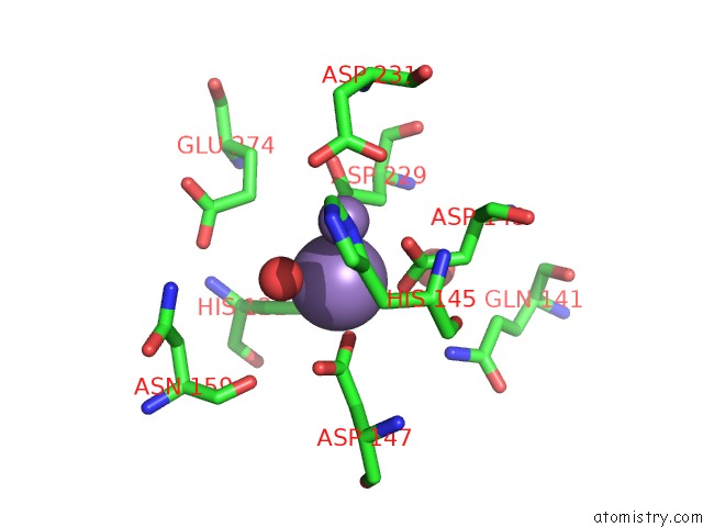
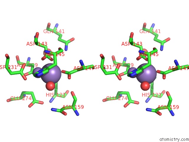
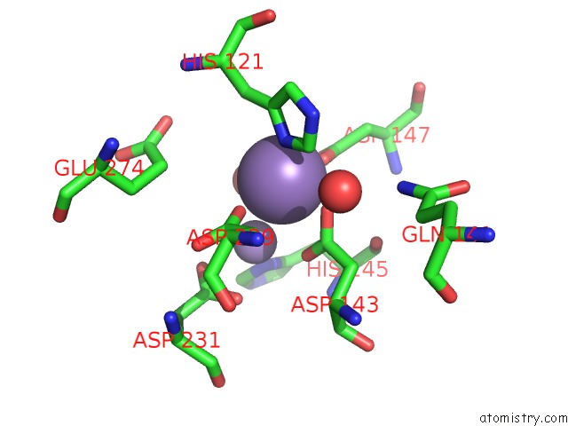
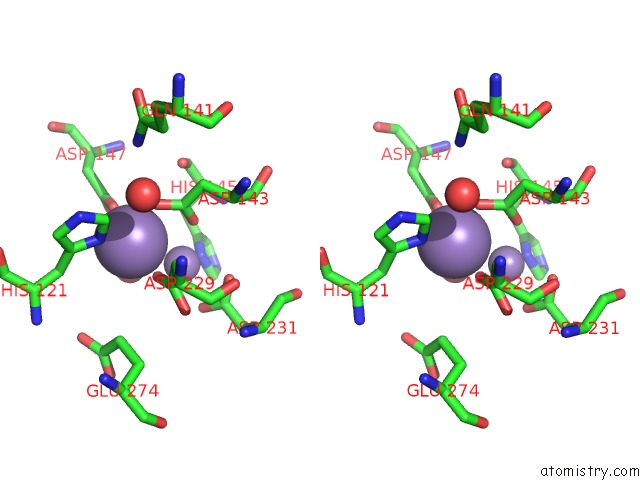
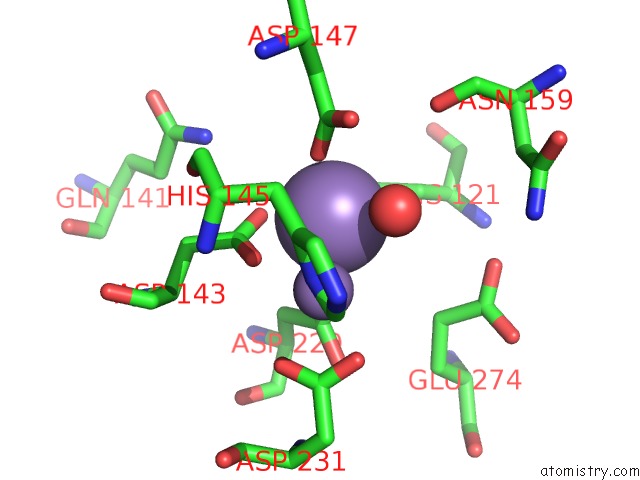
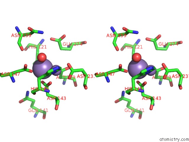
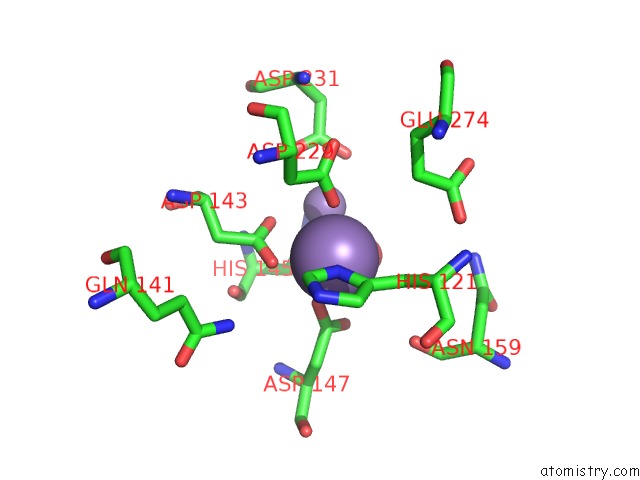
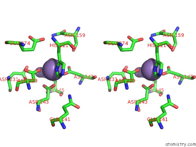
Manganese binding site 1 out of 12 in 1woi
Go back to
Manganese binding site 1 out
of 12 in the Crystal Structure of Agmatinase Reveals Structural Conservation and Inhibition Mechanism of the Ureohydrolase Superfamily
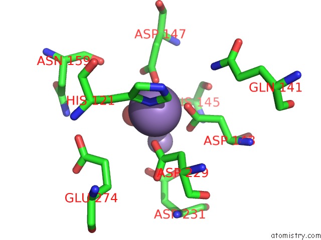
Mono view
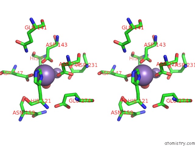
Stereo pair view
Mono view
Stereo pair view
A full contact list of Manganese with other atoms in the Mn binding
site number 1 of Crystal Structure of Agmatinase Reveals Structural Conservation and Inhibition Mechanism of the Ureohydrolase Superfamily within 5.0Å range:
|
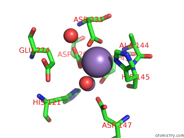
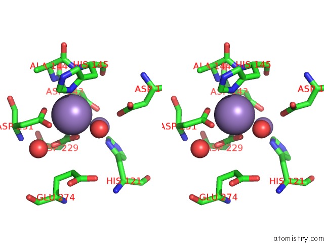
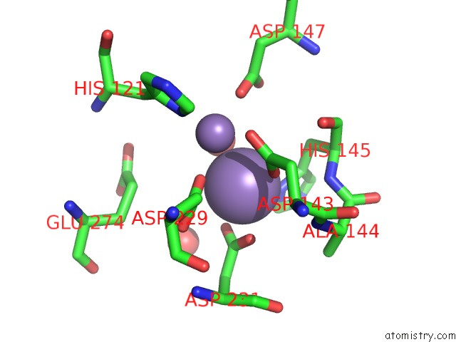
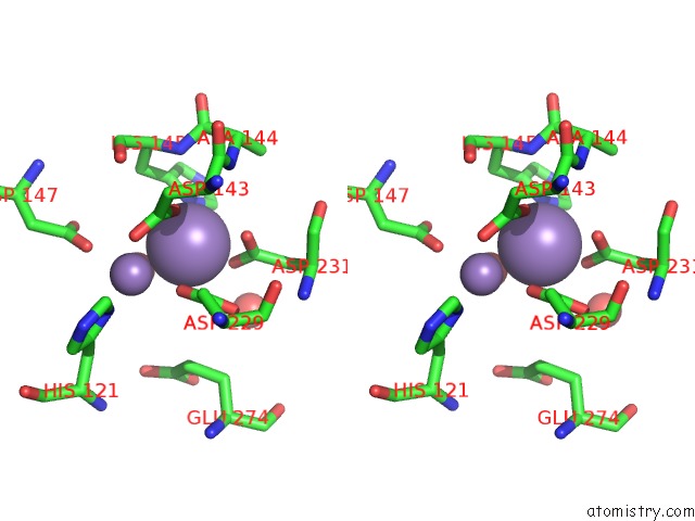
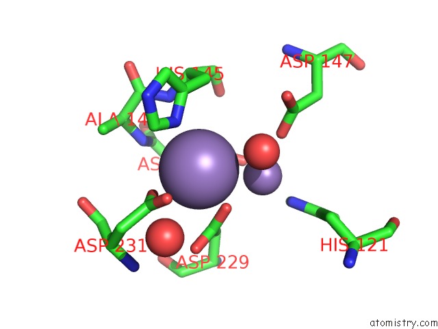
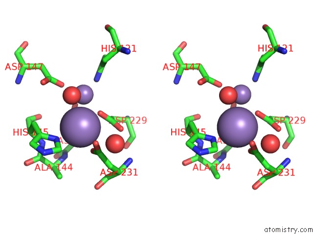
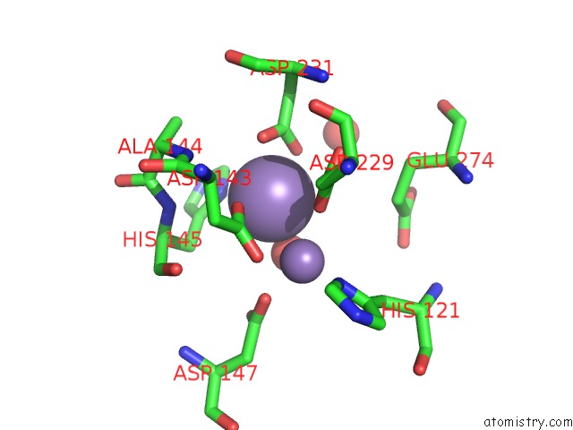
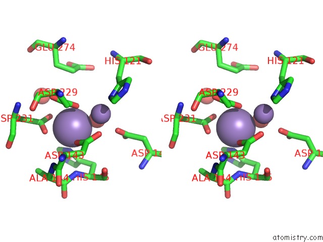
Manganese binding site 2 out of 12 in 1woi
Go back to
Manganese binding site 2 out
of 12 in the Crystal Structure of Agmatinase Reveals Structural Conservation and Inhibition Mechanism of the Ureohydrolase Superfamily
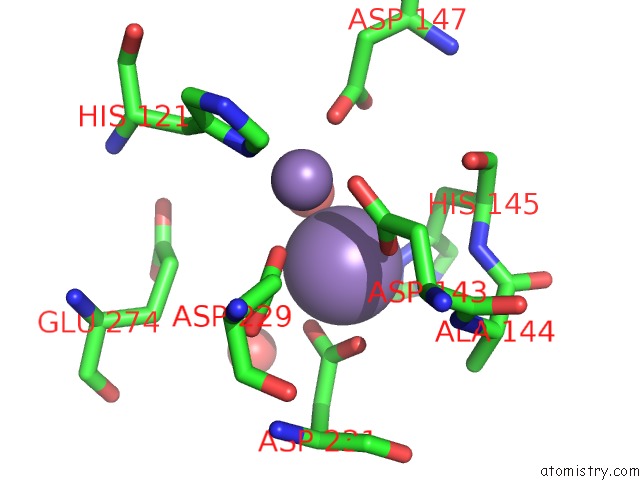
Mono view
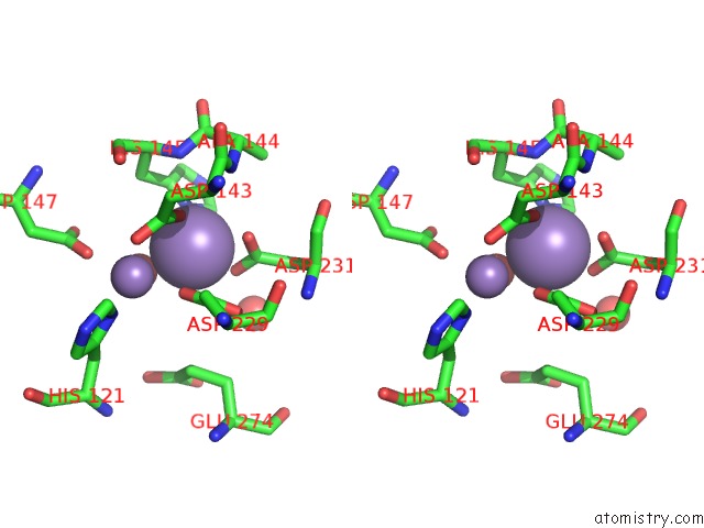
Stereo pair view
Mono view
Stereo pair view
A full contact list of Manganese with other atoms in the Mn binding
site number 2 of Crystal Structure of Agmatinase Reveals Structural Conservation and Inhibition Mechanism of the Ureohydrolase Superfamily within 5.0Å range:
|
Manganese binding site 3 out of 12 in 1woi
Go back to
Manganese binding site 3 out
of 12 in the Crystal Structure of Agmatinase Reveals Structural Conservation and Inhibition Mechanism of the Ureohydrolase Superfamily
Mono view
Stereo pair view
Mono view
Stereo pair view
A full contact list of Manganese with other atoms in the Mn binding
site number 3 of Crystal Structure of Agmatinase Reveals Structural Conservation and Inhibition Mechanism of the Ureohydrolase Superfamily within 5.0Å range:
|
Manganese binding site 4 out of 12 in 1woi
Go back to
Manganese binding site 4 out
of 12 in the Crystal Structure of Agmatinase Reveals Structural Conservation and Inhibition Mechanism of the Ureohydrolase Superfamily
Mono view
Stereo pair view
Mono view
Stereo pair view
A full contact list of Manganese with other atoms in the Mn binding
site number 4 of Crystal Structure of Agmatinase Reveals Structural Conservation and Inhibition Mechanism of the Ureohydrolase Superfamily within 5.0Å range:
|
Manganese binding site 5 out of 12 in 1woi
Go back to
Manganese binding site 5 out
of 12 in the Crystal Structure of Agmatinase Reveals Structural Conservation and Inhibition Mechanism of the Ureohydrolase Superfamily
Mono view
Stereo pair view
Mono view
Stereo pair view
A full contact list of Manganese with other atoms in the Mn binding
site number 5 of Crystal Structure of Agmatinase Reveals Structural Conservation and Inhibition Mechanism of the Ureohydrolase Superfamily within 5.0Å range:
|
Manganese binding site 6 out of 12 in 1woi
Go back to
Manganese binding site 6 out
of 12 in the Crystal Structure of Agmatinase Reveals Structural Conservation and Inhibition Mechanism of the Ureohydrolase Superfamily
Mono view
Stereo pair view
Mono view
Stereo pair view
A full contact list of Manganese with other atoms in the Mn binding
site number 6 of Crystal Structure of Agmatinase Reveals Structural Conservation and Inhibition Mechanism of the Ureohydrolase Superfamily within 5.0Å range:
|
Manganese binding site 7 out of 12 in 1woi
Go back to
Manganese binding site 7 out
of 12 in the Crystal Structure of Agmatinase Reveals Structural Conservation and Inhibition Mechanism of the Ureohydrolase Superfamily
Mono view
Stereo pair view
Mono view
Stereo pair view
A full contact list of Manganese with other atoms in the Mn binding
site number 7 of Crystal Structure of Agmatinase Reveals Structural Conservation and Inhibition Mechanism of the Ureohydrolase Superfamily within 5.0Å range:
|
Manganese binding site 8 out of 12 in 1woi
Go back to
Manganese binding site 8 out
of 12 in the Crystal Structure of Agmatinase Reveals Structural Conservation and Inhibition Mechanism of the Ureohydrolase Superfamily
Mono view
Stereo pair view
Mono view
Stereo pair view
A full contact list of Manganese with other atoms in the Mn binding
site number 8 of Crystal Structure of Agmatinase Reveals Structural Conservation and Inhibition Mechanism of the Ureohydrolase Superfamily within 5.0Å range:
|
Manganese binding site 9 out of 12 in 1woi
Go back to
Manganese binding site 9 out
of 12 in the Crystal Structure of Agmatinase Reveals Structural Conservation and Inhibition Mechanism of the Ureohydrolase Superfamily
Mono view
Stereo pair view
Mono view
Stereo pair view
A full contact list of Manganese with other atoms in the Mn binding
site number 9 of Crystal Structure of Agmatinase Reveals Structural Conservation and Inhibition Mechanism of the Ureohydrolase Superfamily within 5.0Å range:
|
Manganese binding site 10 out of 12 in 1woi
Go back to
Manganese binding site 10 out
of 12 in the Crystal Structure of Agmatinase Reveals Structural Conservation and Inhibition Mechanism of the Ureohydrolase Superfamily
Mono view
Stereo pair view
Mono view
Stereo pair view
A full contact list of Manganese with other atoms in the Mn binding
site number 10 of Crystal Structure of Agmatinase Reveals Structural Conservation and Inhibition Mechanism of the Ureohydrolase Superfamily within 5.0Å range:
|
Reference:
H.J.Ahn,
K.H.Kim,
J.Lee,
J.-Y.Ha,
H.H.Lee,
D.Kim,
H.-J.Yoon,
A.-R.Kwon,
S.W.Suh.
Crystal Structure of Agmatinase Reveals Structural Conservation and Inhibition Mechanism of the Ureohydrolase Superfamily J.Biol.Chem. V. 279 50505 2004.
ISSN: ISSN 0021-9258
PubMed: 15355972
DOI: 10.1074/JBC.M409246200
Page generated: Sat Oct 5 12:56:56 2024
ISSN: ISSN 0021-9258
PubMed: 15355972
DOI: 10.1074/JBC.M409246200
Last articles
Ca in 5N7DCa in 5N5P
Ca in 5N6N
Ca in 5N5K
Ca in 5N5W
Ca in 5N5J
Ca in 5N4L
Ca in 5N3Y
Ca in 5N31
Ca in 5N3V