Manganese »
PDB 1w2c-1xie »
1wbf »
Manganese in PDB 1wbf: Winged Bean Lectin, Saccharide Free Form
Protein crystallography data
The structure of Winged Bean Lectin, Saccharide Free Form, PDB code: 1wbf
was solved by
N.Manoj,
V.R.Srinivas,
K.Suguna,
with X-Ray Crystallography technique. A brief refinement statistics is given in the table below:
| Resolution Low / High (Å) | 8.00 / 2.30 |
| Space group | C 1 2 1 |
| Cell size a, b, c (Å), α, β, γ (°) | 99.159, 75.161, 95.772, 90.00, 106.91, 90.00 |
| R / Rfree (%) | 19.7 / 25.7 |
Other elements in 1wbf:
The structure of Winged Bean Lectin, Saccharide Free Form also contains other interesting chemical elements:
| Calcium | (Ca) | 2 atoms |
Manganese Binding Sites:
The binding sites of Manganese atom in the Winged Bean Lectin, Saccharide Free Form
(pdb code 1wbf). This binding sites where shown within
5.0 Angstroms radius around Manganese atom.
In total 2 binding sites of Manganese where determined in the Winged Bean Lectin, Saccharide Free Form, PDB code: 1wbf:
Jump to Manganese binding site number: 1; 2;
In total 2 binding sites of Manganese where determined in the Winged Bean Lectin, Saccharide Free Form, PDB code: 1wbf:
Jump to Manganese binding site number: 1; 2;
Manganese binding site 1 out of 2 in 1wbf
Go back to
Manganese binding site 1 out
of 2 in the Winged Bean Lectin, Saccharide Free Form
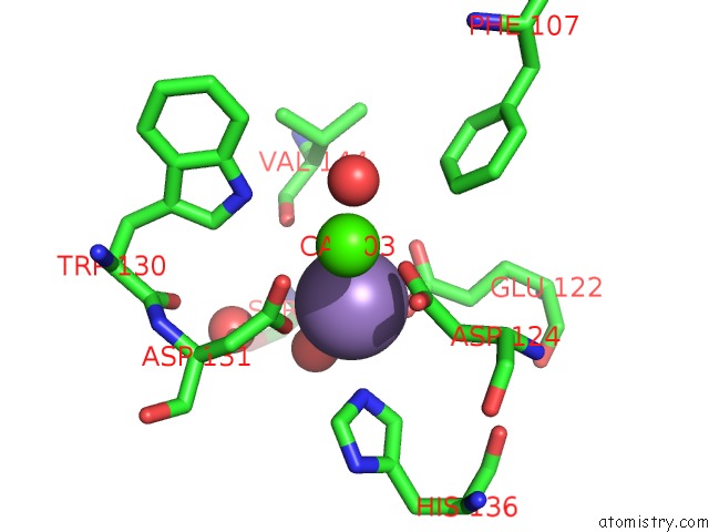
Mono view
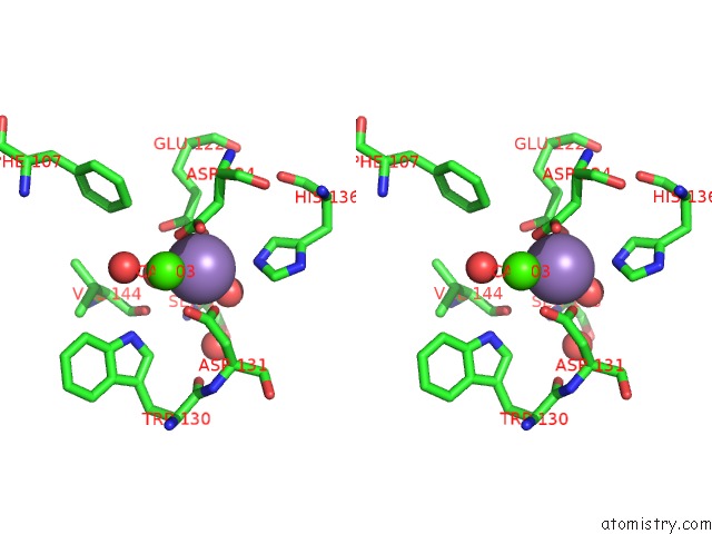
Stereo pair view
Mono view
Stereo pair view
A full contact list of Manganese with other atoms in the Mn binding
site number 1 of Winged Bean Lectin, Saccharide Free Form within 5.0Å range:
|
Manganese binding site 2 out of 2 in 1wbf
Go back to
Manganese binding site 2 out
of 2 in the Winged Bean Lectin, Saccharide Free Form
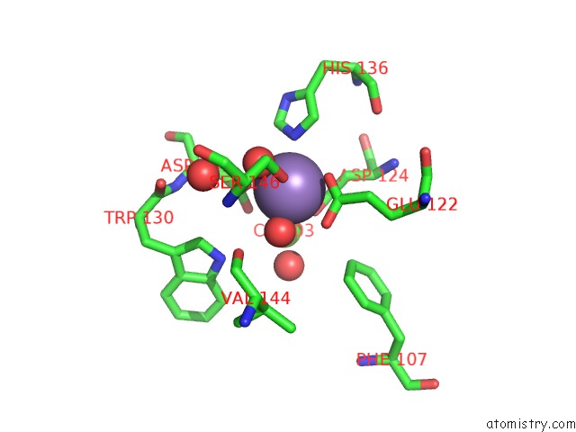
Mono view
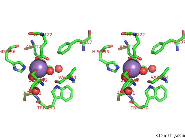
Stereo pair view
Mono view
Stereo pair view
A full contact list of Manganese with other atoms in the Mn binding
site number 2 of Winged Bean Lectin, Saccharide Free Form within 5.0Å range:
|
Reference:
N.Manoj,
V.R.Srinivas,
K.Suguna.
Structure of Basic Winged-Bean Lectin and A Comparison with Its Saccharide-Bound Form. Acta Crystallogr.,Sect.D V. 55 794 1999.
ISSN: ISSN 0907-4449
PubMed: 10089310
DOI: 10.1107/S090744499900044X
Page generated: Sat Oct 5 12:53:15 2024
ISSN: ISSN 0907-4449
PubMed: 10089310
DOI: 10.1107/S090744499900044X
Last articles
Zn in 9MJ5Zn in 9HNW
Zn in 9G0L
Zn in 9FNE
Zn in 9DZN
Zn in 9E0I
Zn in 9D32
Zn in 9DAK
Zn in 8ZXC
Zn in 8ZUF