Manganese »
PDB 1uvj-1vzx »
1vby »
Manganese in PDB 1vby: Crystal Structure of the Hepatitis Delta Virus Gemonic Ribozyme Precursor, with C75U Mutaion, and MN2+ Bound
Protein crystallography data
The structure of Crystal Structure of the Hepatitis Delta Virus Gemonic Ribozyme Precursor, with C75U Mutaion, and MN2+ Bound, PDB code: 1vby
was solved by
A.Ke,
K.Zhou,
F.Ding,
J.H.D.Cate,
J.A.Doudna,
with X-Ray Crystallography technique. A brief refinement statistics is given in the table below:
| Resolution Low / High (Å) | 42.16 / 2.90 |
| Space group | H 3 2 |
| Cell size a, b, c (Å), α, β, γ (°) | 108.589, 108.589, 190.519, 90.00, 90.00, 120.00 |
| R / Rfree (%) | 23.9 / 28.4 |
Other elements in 1vby:
The structure of Crystal Structure of the Hepatitis Delta Virus Gemonic Ribozyme Precursor, with C75U Mutaion, and MN2+ Bound also contains other interesting chemical elements:
| Sodium | (Na) | 1 atom |
Manganese Binding Sites:
The binding sites of Manganese atom in the Crystal Structure of the Hepatitis Delta Virus Gemonic Ribozyme Precursor, with C75U Mutaion, and MN2+ Bound
(pdb code 1vby). This binding sites where shown within
5.0 Angstroms radius around Manganese atom.
In total 2 binding sites of Manganese where determined in the Crystal Structure of the Hepatitis Delta Virus Gemonic Ribozyme Precursor, with C75U Mutaion, and MN2+ Bound, PDB code: 1vby:
Jump to Manganese binding site number: 1; 2;
In total 2 binding sites of Manganese where determined in the Crystal Structure of the Hepatitis Delta Virus Gemonic Ribozyme Precursor, with C75U Mutaion, and MN2+ Bound, PDB code: 1vby:
Jump to Manganese binding site number: 1; 2;
Manganese binding site 1 out of 2 in 1vby
Go back to
Manganese binding site 1 out
of 2 in the Crystal Structure of the Hepatitis Delta Virus Gemonic Ribozyme Precursor, with C75U Mutaion, and MN2+ Bound
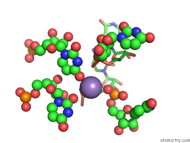
Mono view
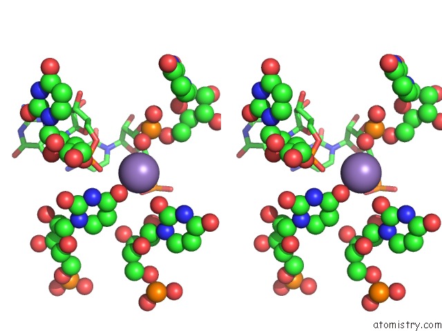
Stereo pair view
Mono view
Stereo pair view
A full contact list of Manganese with other atoms in the Mn binding
site number 1 of Crystal Structure of the Hepatitis Delta Virus Gemonic Ribozyme Precursor, with C75U Mutaion, and MN2+ Bound within 5.0Å range:
|
Manganese binding site 2 out of 2 in 1vby
Go back to
Manganese binding site 2 out
of 2 in the Crystal Structure of the Hepatitis Delta Virus Gemonic Ribozyme Precursor, with C75U Mutaion, and MN2+ Bound
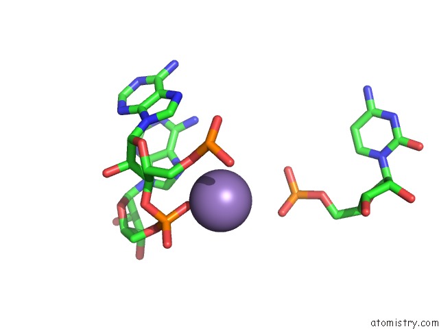
Mono view
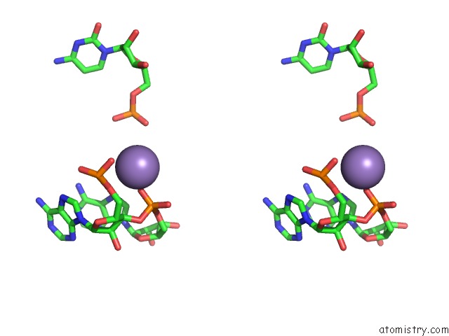
Stereo pair view
Mono view
Stereo pair view
A full contact list of Manganese with other atoms in the Mn binding
site number 2 of Crystal Structure of the Hepatitis Delta Virus Gemonic Ribozyme Precursor, with C75U Mutaion, and MN2+ Bound within 5.0Å range:
|
Reference:
A.Ke,
K.Zhou,
F.Ding,
J.H.D.Cate,
J.A.Doudna.
A Conformational Switch Controls Hepatitis Delta Virus Ribozyme Catalysis Nature V. 429 201 2004.
ISSN: ISSN 0028-0836
PubMed: 15141216
DOI: 10.1038/NATURE02522
Page generated: Sat Oct 5 12:47:13 2024
ISSN: ISSN 0028-0836
PubMed: 15141216
DOI: 10.1038/NATURE02522
Last articles
Zn in 9MJ5Zn in 9HNW
Zn in 9G0L
Zn in 9FNE
Zn in 9DZN
Zn in 9E0I
Zn in 9D32
Zn in 9DAK
Zn in 8ZXC
Zn in 8ZUF