Manganese »
PDB 1uvj-1vzx »
1v9q »
Manganese in PDB 1v9q: Crystal Structure of An Artificial Metalloprotein:Mn(III)(3,3'-ME2- Salophen)/Apo-A71G Myoglobin
Protein crystallography data
The structure of Crystal Structure of An Artificial Metalloprotein:Mn(III)(3,3'-ME2- Salophen)/Apo-A71G Myoglobin, PDB code: 1v9q
was solved by
T.Ueno,
T.Koshiyama,
M.Kono,
K.Kondo,
M.Ohashi,
A.Suzuki,
T.Yamane,
Y.Watanabe,
with X-Ray Crystallography technique. A brief refinement statistics is given in the table below:
| Resolution Low / High (Å) | 27.01 / 1.45 |
| Space group | P 21 21 21 |
| Cell size a, b, c (Å), α, β, γ (°) | 33.284, 57.848, 75.418, 90.00, 90.00, 90.00 |
| R / Rfree (%) | 20.6 / 21.8 |
Manganese Binding Sites:
The binding sites of Manganese atom in the Crystal Structure of An Artificial Metalloprotein:Mn(III)(3,3'-ME2- Salophen)/Apo-A71G Myoglobin
(pdb code 1v9q). This binding sites where shown within
5.0 Angstroms radius around Manganese atom.
In total only one binding site of Manganese was determined in the Crystal Structure of An Artificial Metalloprotein:Mn(III)(3,3'-ME2- Salophen)/Apo-A71G Myoglobin, PDB code: 1v9q:
In total only one binding site of Manganese was determined in the Crystal Structure of An Artificial Metalloprotein:Mn(III)(3,3'-ME2- Salophen)/Apo-A71G Myoglobin, PDB code: 1v9q:
Manganese binding site 1 out of 1 in 1v9q
Go back to
Manganese binding site 1 out
of 1 in the Crystal Structure of An Artificial Metalloprotein:Mn(III)(3,3'-ME2- Salophen)/Apo-A71G Myoglobin
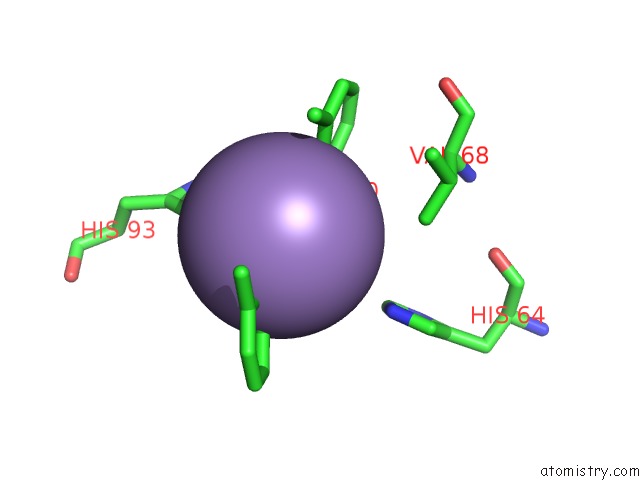
Mono view
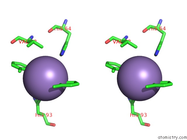
Stereo pair view
Mono view
Stereo pair view
A full contact list of Manganese with other atoms in the Mn binding
site number 1 of Crystal Structure of An Artificial Metalloprotein:Mn(III)(3,3'-ME2- Salophen)/Apo-A71G Myoglobin within 5.0Å range:
|
Reference:
T.Ueno,
T.Koshiyama,
M.Ohashi,
K.Kondo,
M.Kono,
A.Suzuki,
T.Yamane,
Y.Watanabe.
Coordinated Design of Cofactor and Active Site Structures in Development of New Protein Catalysts J.Am.Chem.Soc. V. 127 6556 2005.
ISSN: ISSN 0002-7863
PubMed: 15869276
DOI: 10.1021/JA045995Q
Page generated: Sat Oct 5 12:46:00 2024
ISSN: ISSN 0002-7863
PubMed: 15869276
DOI: 10.1021/JA045995Q
Last articles
Zn in 9J0NZn in 9J0O
Zn in 9J0P
Zn in 9FJX
Zn in 9EKB
Zn in 9C0F
Zn in 9CAH
Zn in 9CH0
Zn in 9CH3
Zn in 9CH1