Manganese »
PDB 1uvj-1vzx »
1v83 »
Manganese in PDB 1v83: Crystal Structure of Human Glcat-P in Complex with Udp and MN2+
Enzymatic activity of Crystal Structure of Human Glcat-P in Complex with Udp and MN2+
All present enzymatic activity of Crystal Structure of Human Glcat-P in Complex with Udp and MN2+:
2.4.1.135;
2.4.1.135;
Protein crystallography data
The structure of Crystal Structure of Human Glcat-P in Complex with Udp and MN2+, PDB code: 1v83
was solved by
S.Kakuda,
T.Shiba,
M.Ishiguro,
H.Tagawa,
S.Oka,
Y.Kajihara,
T.Kawasaki,
S.Wakatsuki,
R.Kato,
with X-Ray Crystallography technique. A brief refinement statistics is given in the table below:
| Resolution Low / High (Å) | 40.00 / 1.90 |
| Space group | P 21 21 21 |
| Cell size a, b, c (Å), α, β, γ (°) | 61.737, 85.701, 122.911, 90.00, 90.00, 90.00 |
| R / Rfree (%) | 20.1 / 22.9 |
Manganese Binding Sites:
The binding sites of Manganese atom in the Crystal Structure of Human Glcat-P in Complex with Udp and MN2+
(pdb code 1v83). This binding sites where shown within
5.0 Angstroms radius around Manganese atom.
In total 2 binding sites of Manganese where determined in the Crystal Structure of Human Glcat-P in Complex with Udp and MN2+, PDB code: 1v83:
Jump to Manganese binding site number: 1; 2;
In total 2 binding sites of Manganese where determined in the Crystal Structure of Human Glcat-P in Complex with Udp and MN2+, PDB code: 1v83:
Jump to Manganese binding site number: 1; 2;
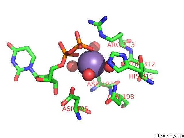
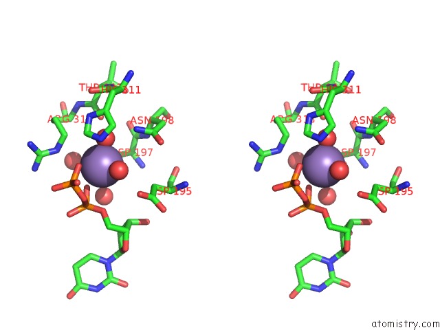
Manganese binding site 1 out of 2 in 1v83
Go back to
Manganese binding site 1 out
of 2 in the Crystal Structure of Human Glcat-P in Complex with Udp and MN2+

Mono view

Stereo pair view
Mono view
Stereo pair view
A full contact list of Manganese with other atoms in the Mn binding
site number 1 of Crystal Structure of Human Glcat-P in Complex with Udp and MN2+ within 5.0Å range:
|
Manganese binding site 2 out of 2 in 1v83
Go back to
Manganese binding site 2 out
of 2 in the Crystal Structure of Human Glcat-P in Complex with Udp and MN2+
Mono view
Stereo pair view
Mono view
Stereo pair view
A full contact list of Manganese with other atoms in the Mn binding
site number 2 of Crystal Structure of Human Glcat-P in Complex with Udp and MN2+ within 5.0Å range:
|
Reference:
S.Kakuda,
T.Shiba,
M.Ishiguro,
H.Tagawa,
S.Oka,
Y.Kajihara,
T.Kawasaki,
S.Wakatsuki,
R.Kato.
Structural Basis For Acceptor Substrate Recognition of A Human Glucuronyltransferase, Glcat-P, An Enzyme Critical in the Biosynthesis of the Carbohydrate Epitope Hnk-1 J.Biol.Chem. V. 279 22693 2004.
ISSN: ISSN 0021-9258
PubMed: 14993226
DOI: 10.1074/JBC.M400622200
Page generated: Sat Oct 5 12:45:53 2024
ISSN: ISSN 0021-9258
PubMed: 14993226
DOI: 10.1074/JBC.M400622200
Last articles
Zn in 9MJ5Zn in 9HNW
Zn in 9G0L
Zn in 9FNE
Zn in 9DZN
Zn in 9E0I
Zn in 9D32
Zn in 9DAK
Zn in 8ZXC
Zn in 8ZUF