Manganese »
PDB 1uvj-1vzx »
1v6j »
Manganese in PDB 1v6j: Peanut Lectin-Lactose Complex Crystallized in Orthorhombic Form at Acidic pH
Protein crystallography data
The structure of Peanut Lectin-Lactose Complex Crystallized in Orthorhombic Form at Acidic pH, PDB code: 1v6j
was solved by
S.Kundhavai Natchiar,
A.Arockia Jeyaprakash,
T.N.C.Ramya,
C.J.Thomas,
K.Suguna,
A.Surolia,
M.Vijayan,
with X-Ray Crystallography technique. A brief refinement statistics is given in the table below:
| Resolution Low / High (Å) | 20.00 / 2.90 |
| Space group | P 21 21 2 |
| Cell size a, b, c (Å), α, β, γ (°) | 128.733, 125.531, 76.050, 90.00, 90.00, 90.00 |
| R / Rfree (%) | 16.1 / 22 |
Other elements in 1v6j:
The structure of Peanut Lectin-Lactose Complex Crystallized in Orthorhombic Form at Acidic pH also contains other interesting chemical elements:
| Calcium | (Ca) | 4 atoms |
Manganese Binding Sites:
The binding sites of Manganese atom in the Peanut Lectin-Lactose Complex Crystallized in Orthorhombic Form at Acidic pH
(pdb code 1v6j). This binding sites where shown within
5.0 Angstroms radius around Manganese atom.
In total 4 binding sites of Manganese where determined in the Peanut Lectin-Lactose Complex Crystallized in Orthorhombic Form at Acidic pH, PDB code: 1v6j:
Jump to Manganese binding site number: 1; 2; 3; 4;
In total 4 binding sites of Manganese where determined in the Peanut Lectin-Lactose Complex Crystallized in Orthorhombic Form at Acidic pH, PDB code: 1v6j:
Jump to Manganese binding site number: 1; 2; 3; 4;
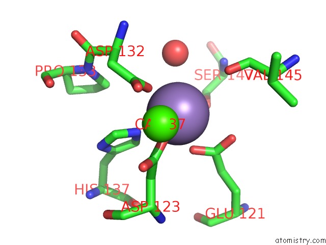
Manganese binding site 1 out of 4 in 1v6j
Go back to
Manganese binding site 1 out
of 4 in the Peanut Lectin-Lactose Complex Crystallized in Orthorhombic Form at Acidic pH

Mono view

Stereo pair view
Mono view
Stereo pair view
A full contact list of Manganese with other atoms in the Mn binding
site number 1 of Peanut Lectin-Lactose Complex Crystallized in Orthorhombic Form at Acidic pH within 5.0Å range:
|
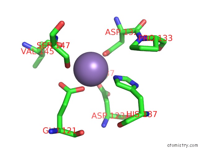
Manganese binding site 2 out of 4 in 1v6j
Go back to
Manganese binding site 2 out
of 4 in the Peanut Lectin-Lactose Complex Crystallized in Orthorhombic Form at Acidic pH
Mono view

Stereo pair view
Mono view
Stereo pair view
A full contact list of Manganese with other atoms in the Mn binding
site number 2 of Peanut Lectin-Lactose Complex Crystallized in Orthorhombic Form at Acidic pH within 5.0Å range:
|
Manganese binding site 3 out of 4 in 1v6j
Go back to
Manganese binding site 3 out
of 4 in the Peanut Lectin-Lactose Complex Crystallized in Orthorhombic Form at Acidic pH
Mono view
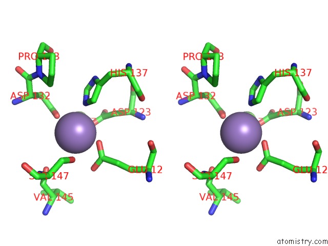
Stereo pair view
Mono view
Stereo pair view
A full contact list of Manganese with other atoms in the Mn binding
site number 3 of Peanut Lectin-Lactose Complex Crystallized in Orthorhombic Form at Acidic pH within 5.0Å range:
|
Manganese binding site 4 out of 4 in 1v6j
Go back to
Manganese binding site 4 out
of 4 in the Peanut Lectin-Lactose Complex Crystallized in Orthorhombic Form at Acidic pH

Mono view

Stereo pair view
Mono view
Stereo pair view
A full contact list of Manganese with other atoms in the Mn binding
site number 4 of Peanut Lectin-Lactose Complex Crystallized in Orthorhombic Form at Acidic pH within 5.0Å range:
|
Reference:
S.Kundhavai Natchiar,
A.Arockia Jeyaprakash,
T.N.Ramya,
C.J.Thomas,
K.Suguna,
A.Surolia,
M.Vijayan.
Structural Plasticity of Peanut Lectin: An X-Ray Analysis Involving Variation in pH, Ligand Binding and Crystal Structure. Acta Crystallogr.,Sect.D V. 60 211 2004.
ISSN: ISSN 0907-4449
PubMed: 14747696
DOI: 10.1107/S090744490302849X
Page generated: Sat Oct 5 12:42:54 2024
ISSN: ISSN 0907-4449
PubMed: 14747696
DOI: 10.1107/S090744490302849X
Last articles
F in 7K1HF in 7K2U
F in 7K1F
F in 7K1E
F in 7K1I
F in 7K1D
F in 7JY4
F in 7K0V
F in 7JYM
F in 7K15