Manganese »
PDB 1ss9-1uvi »
1ucg »
Manganese in PDB 1ucg: Crystal Structure of Ribonuclease MC1 N71T Mutant
Enzymatic activity of Crystal Structure of Ribonuclease MC1 N71T Mutant
All present enzymatic activity of Crystal Structure of Ribonuclease MC1 N71T Mutant:
3.1.27.1;
3.1.27.1;
Protein crystallography data
The structure of Crystal Structure of Ribonuclease MC1 N71T Mutant, PDB code: 1ucg
was solved by
A.Suzuki,
T.Numata,
M.Yao,
I.Tanaka,
M.Kimura,
with X-Ray Crystallography technique. A brief refinement statistics is given in the table below:
| Resolution Low / High (Å) | 10.00 / 1.65 |
| Space group | P 21 21 2 |
| Cell size a, b, c (Å), α, β, γ (°) | 58.790, 135.900, 52.390, 90.00, 90.00, 90.00 |
| R / Rfree (%) | 18.1 / 19.6 |
Manganese Binding Sites:
The binding sites of Manganese atom in the Crystal Structure of Ribonuclease MC1 N71T Mutant
(pdb code 1ucg). This binding sites where shown within
5.0 Angstroms radius around Manganese atom.
In total 4 binding sites of Manganese where determined in the Crystal Structure of Ribonuclease MC1 N71T Mutant, PDB code: 1ucg:
Jump to Manganese binding site number: 1; 2; 3; 4;
In total 4 binding sites of Manganese where determined in the Crystal Structure of Ribonuclease MC1 N71T Mutant, PDB code: 1ucg:
Jump to Manganese binding site number: 1; 2; 3; 4;
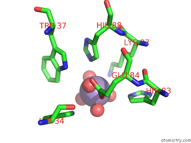
Manganese binding site 1 out of 4 in 1ucg
Go back to
Manganese binding site 1 out
of 4 in the Crystal Structure of Ribonuclease MC1 N71T Mutant
Mono view

Stereo pair view
Mono view
Stereo pair view
A full contact list of Manganese with other atoms in the Mn binding
site number 1 of Crystal Structure of Ribonuclease MC1 N71T Mutant within 5.0Å range:
|
Manganese binding site 2 out of 4 in 1ucg
Go back to
Manganese binding site 2 out
of 4 in the Crystal Structure of Ribonuclease MC1 N71T Mutant

Mono view

Stereo pair view
Mono view
Stereo pair view
A full contact list of Manganese with other atoms in the Mn binding
site number 2 of Crystal Structure of Ribonuclease MC1 N71T Mutant within 5.0Å range:
|
Manganese binding site 3 out of 4 in 1ucg
Go back to
Manganese binding site 3 out
of 4 in the Crystal Structure of Ribonuclease MC1 N71T Mutant

Mono view

Stereo pair view
Mono view
Stereo pair view
A full contact list of Manganese with other atoms in the Mn binding
site number 3 of Crystal Structure of Ribonuclease MC1 N71T Mutant within 5.0Å range:
|
Manganese binding site 4 out of 4 in 1ucg
Go back to
Manganese binding site 4 out
of 4 in the Crystal Structure of Ribonuclease MC1 N71T Mutant

Mono view

Stereo pair view
Mono view
Stereo pair view
A full contact list of Manganese with other atoms in the Mn binding
site number 4 of Crystal Structure of Ribonuclease MC1 N71T Mutant within 5.0Å range:
|
Reference:
T.Numata,
A.Suzuki,
Y.Kakuta,
K.Kimura,
M.Yao,
I.Tanaka,
Y.Yoshida,
T.Ueda,
M.Kimura.
Crystal Structures of the Ribonuclease MC1 Mutants N71T and N71S in Complex with 5'-Gmp: Structural Basis For Alterations in Substrate Specificity Biochemistry V. 42 5270 2003.
ISSN: ISSN 0006-2960
PubMed: 12731868
DOI: 10.1021/BI034103G
Page generated: Sat Oct 5 12:38:06 2024
ISSN: ISSN 0006-2960
PubMed: 12731868
DOI: 10.1021/BI034103G
Last articles
F in 7LZWF in 7LY8
F in 7LWG
F in 7LZV
F in 7LZF
F in 7LZD
F in 7LZA
F in 7LVX
F in 7LUN
F in 7LVR