Manganese »
PDB 1ss9-1uvi »
1tc2 »
Manganese in PDB 1tc2: Ternary Substrate Complex of the Hypoxanthine Phosphoribosyltransferase From Trypanosoma Cruzi
Enzymatic activity of Ternary Substrate Complex of the Hypoxanthine Phosphoribosyltransferase From Trypanosoma Cruzi
All present enzymatic activity of Ternary Substrate Complex of the Hypoxanthine Phosphoribosyltransferase From Trypanosoma Cruzi:
2.4.2.8;
2.4.2.8;
Protein crystallography data
The structure of Ternary Substrate Complex of the Hypoxanthine Phosphoribosyltransferase From Trypanosoma Cruzi, PDB code: 1tc2
was solved by
P.J.Focia,
S.P.Craig Iii,
A.E.Eakin,
with X-Ray Crystallography technique. A brief refinement statistics is given in the table below:
| Resolution Low / High (Å) | 6.00 / 1.81 |
| Space group | P 1 21 1 |
| Cell size a, b, c (Å), α, β, γ (°) | 39.420, 101.760, 51.920, 90.00, 94.23, 90.00 |
| R / Rfree (%) | 18.3 / 23.2 |
Other elements in 1tc2:
The structure of Ternary Substrate Complex of the Hypoxanthine Phosphoribosyltransferase From Trypanosoma Cruzi also contains other interesting chemical elements:
| Magnesium | (Mg) | 3 atoms |
Manganese Binding Sites:
The binding sites of Manganese atom in the Ternary Substrate Complex of the Hypoxanthine Phosphoribosyltransferase From Trypanosoma Cruzi
(pdb code 1tc2). This binding sites where shown within
5.0 Angstroms radius around Manganese atom.
In total only one binding site of Manganese was determined in the Ternary Substrate Complex of the Hypoxanthine Phosphoribosyltransferase From Trypanosoma Cruzi, PDB code: 1tc2:
In total only one binding site of Manganese was determined in the Ternary Substrate Complex of the Hypoxanthine Phosphoribosyltransferase From Trypanosoma Cruzi, PDB code: 1tc2:
Manganese binding site 1 out of 1 in 1tc2
Go back to
Manganese binding site 1 out
of 1 in the Ternary Substrate Complex of the Hypoxanthine Phosphoribosyltransferase From Trypanosoma Cruzi
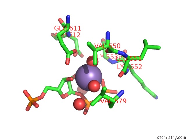
Mono view
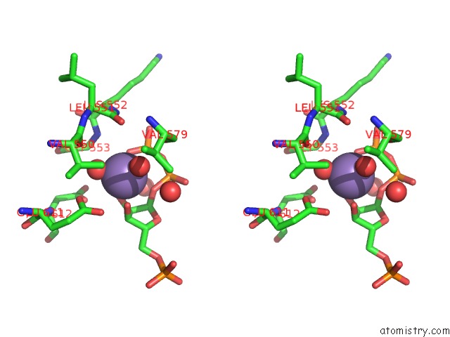
Stereo pair view
Mono view
Stereo pair view
A full contact list of Manganese with other atoms in the Mn binding
site number 1 of Ternary Substrate Complex of the Hypoxanthine Phosphoribosyltransferase From Trypanosoma Cruzi within 5.0Å range:
|
Reference:
P.J.Focia,
S.P.Craig Iii,
A.E.Eakin.
Approaching the Transition State in the Crystal Structure of A Phosphoribosyltransferase. Biochemistry V. 37 17120 1998.
ISSN: ISSN 0006-2960
PubMed: 9860824
DOI: 10.1021/BI9821465
Page generated: Sat Oct 5 12:33:31 2024
ISSN: ISSN 0006-2960
PubMed: 9860824
DOI: 10.1021/BI9821465
Last articles
Zn in 9J0NZn in 9J0O
Zn in 9J0P
Zn in 9FJX
Zn in 9EKB
Zn in 9C0F
Zn in 9CAH
Zn in 9CH0
Zn in 9CH3
Zn in 9CH1